Comparative Study
. 2006 May 3;26(18):4949-55.
doi: 10.1523/JNEUROSCI.4573-05.2006.

Involvement of protein synthesis and degradation in long-term potentiation of Schaffer collateral CA1 synapses

Affiliations
Comparative Study

Involvement of protein synthesis and degradation in long-term potentiation of Schaffer collateral CA1 synapses

Anna Karpova et al. J Neurosci. .

Abstract

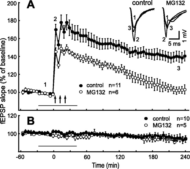
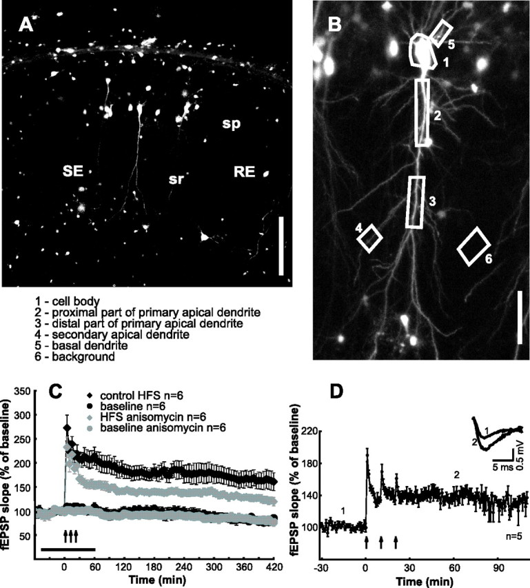
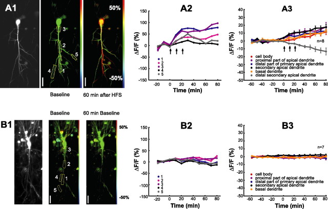
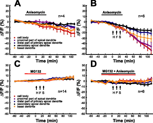
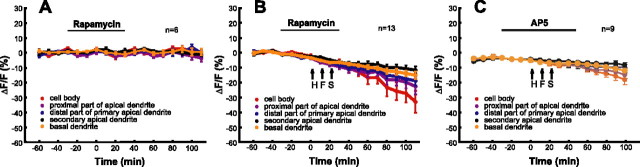
Expression of synaptic plasticity involves the translation of mRNA into protein and, probably, active protein degradation via the proteasome pathway. Here, we report on the rapid activation of synthesis and degradation of a probe protein with the induction of long-term potentiation (LTP) in the hippocampal Schaffer collateral CA1 pathway. The proteasome inhibitor MG132 significantly reduced the field EPSP slope potentiation and LTP maintenance without acutely affecting basal synaptic transmission. To visualize protein dynamics, CA1 pyramidal cells of hippocampal slices were transfected with Semliki Forest virus particles expressing a recombinant RNA. This RNA contained the coding sequence for a degradable green fluorescence protein with a nuclear localization signal (NLS-d1EGFP) followed by a 3'- untranslated region dendritic targeting sequence. NLS-d1EGFP fluorescence remained stable in the low-frequency test stimulation but increased with LTP induction in the cell body and in most dendritic compartments of CA1 neurons. Applying anisomycin, a protein synthesis inhibitor, caused NLS-d1EGFP levels to decline; a proteasome inhibitor MG132 reversed this effect. In the presence of anisomycin, LTP induction accelerated the degradation of NLS-d1EGFP. When both inhibitors were present, NLS-d1EGFP levels remained unaffected by LTP induction. Moreover, LTP-induced acceleration of NLS-d1EGFP synthesis was blocked by rapamycin, which is consistent with the involvement of dendritic mammalian target of rapamycin in LTP-triggered translational activity. Our results clearly demonstrate that LTP induction not only leads to a rapid increase in the rate of protein synthesis but also accelerates protein degradation via the proteasome system.


Figures

Figure 1.
Figure 1.
Inhibition of proteasome activity decreases the magnitude of short-term and late-phase LTP in hippocampal slices. A, Shown are ensemble averages for experiments with HFS in control ACSF (filled circles; n = 11) and in the presence of 10 μm MG132 (n = 6). HFS consisted of three 100 Hz trains delivered at 10 min intervals after time point zero. The inset shows representative fEPSPs for the time points indicated. B, Baseline recordings in control ACSF (filled circles; n = 10) and in the presence of 10 μm MG132 (n = 5). The arrows indicate the times of HFS, and the horizontal bar represents the time of drug application.
Figure 2.
Figure 2.
LTP in hippocampal slices transfected with reporter construct. A, Fluorescence image of CA1 region of hippocampal slice 18 h after transfection. The CA1 pyramidal cells in stratum pyramidale (sp) can be identified by basal and apical dendrites. Stimulation (SE) and recording (RE) pipettes are localized in stratum radiatum (sr). Scale bar, 300 μm. B, Z-projection image of a single CA1 neuron with description of ROIs (see A) used for analysis. Scale bar, 85 μm. C, Stable baseline recordings, expression of long-lasting LTP, and the effect of anisomycin on LTP maintenance in slices maintained for >24 h. Ensemble averages for experiments with HFS (n = 6), baseline recordings (n = 6), HFS in the presence of anisomycin (n = 6; horizontal bar), and baseline in the presence of anisomycin (n = 6) are shown. D, Electrophysiological recordings in transfected slices. Ensemble averages for experiments with HFS (n = 5) are shown. The inset shows representative fEPSPs for the time points indicated.
Figure 3.
Figure 3.
HFS activates synthesis of reporter protein. A1, B1, Fluorescence images of pyramidal cells transfected with reporter construct. Left baseline fluorescence, middle, and right color-coded changes from baseline fluorescence before (middle) and 60 min after (right) HFS (A1) or with continued baseline recordings (B1). Colors indicate the following: decrease, blue; no change, green; increase, red. Scale from −50 to +50%. A2, B2, Time course of fluorescence levels in ROIs from a cell subjected to HFS (A2) and a control cell without HFS (B2). ROIs are indicated in A1 and B1. A3, B3, Ensemble average of reporter protein levels from ROIs as defined in Figure 2B. Signals from secondary apical dendrites (distant from the stimulation electrode) that exhibited decreasing fluorescence levels after HFS were pooled together and represented by gray symbols in A3. Scale bars, 70 μm.
Figure 4.
Figure 4.
HFS activates protein degradation. A–F, The diagrams show ensemble averages of the time course of reporter protein levels under different experimental conditions. Horizontal bars indicate drug application times. A, Anisomycin (25 μm; n = 4). B, HFS in the presence of anisomycin. C, HFS in the presence of MG132 (10 μm). D, HFS in the presence of MG132 and anisomycin.
Figure 5.
Figure 5.
Inhibition of translation by rapamycin and prevention of LTP expression by AP-5 prevent HFS-induced protein synthesis but not protein degradation. A, Rapamycin (200 nm; n = 6) applied during baseline recordings. B, HFS in the presence of rapamycin (n = 13). C, HFS in the presence of AP-5 (n = 9).

References

    1. Aakalu G, Smith WB, Nguyen N, Jiang C, Schuman EM (2001). Dynamic visualization of local protein synthesis in hippocampal neurons. Neuron 30:489–502. - PubMed
    1. Behnisch T, Matsushita S, Knöpfel T (2004). Imaging of gene expression during long-term potentiation. NeuroReport 15:2039–2043. - PubMed
    1. Bingol B, Schuman EM (2004). A proteasome-sensitive connection between PSD-95 and GluR1 endocytosis. Neuropharmacology 47:755–763. - PubMed
    1. Blichenberg A, Schwanke B, Rehbein M, Garner CC, Richter D, Kindler S (1999). Identification of a cis-acting dendritic targeting element in MAP2 mRNAs. J Neurosci 19:8818–8829. - PMC - PubMed
    1. Bliss TV, Collingridge GL (1993). A synaptic model of memory: long-term potentiation in the hippocampus. Nature 361:31–39. - PubMed

Publication types

MeSH terms

LinkOut - more resources