. 2006 Jul;75(1):56-67.
doi: 10.1095/biolreprod.105.047225. Epub 2006 May 3.

Vascular endothelial growth factor and kinase domain region receptor are involved in both seminiferous cord formation and vascular development during testis morphogenesis in the rat

Affiliations

Vascular endothelial growth factor and kinase domain region receptor are involved in both seminiferous cord formation and vascular development during testis morphogenesis in the rat

Rebecca C Bott et al. Biol Reprod. 2006 Jul.

Abstract

Morphological male sex determination is dependent on migration of endothelial and preperitubular cells from the adjacent mesonephros into the developing testis. Our hypothesis is that VEGFA and its receptor KDR are necessary for both testicular cord formation and neovascularization. The Vegfa gene has 8 exons with many splice variants. Vegfa120, Vegfa164, and Vegfa188 mRNA isoforms were detected on Embryonic Day (E) 13.5 (plug date=E0) in the rat. Vegfa120, Vegfa144, Vegfa164, Vegfa188, and Vegfa205 mRNA were detected at E18 and Postnatal Day 3 (P3). Kdr mRNA was present on E13.5, whereas Fms-like tyrosine kinase 1 receptor (Flt1) mRNA was not detected until E18. VEGFA protein was localized to Sertoli cells at cord formation and KDR to germ and interstitial cells. The VEGFA signaling inhibitors SU1498 (40 microM) and VEGFR-TKI (8 microM) inhibited cord formation in E13 testis cultures with 90% reduced vascular density (P<0.01) in VEGFR-TKI-treated organs. Furthermore, Je-11 (10 microM), an antagonist to VEGFA, also perturbed cord formation and inhibited vascular density by more than 50% (P<0.01). To determine signal transduction pathways involved in VEGFA's regulation of testis morphogenesis, E13 testis were treated with LY 294002 (15 microM), a phosphoinositide 3-kinase (PI3K) pathway inhibitor, resulting in inhibition of both vascular density (46%) and cord formation. Thus, we support our hypothesis and conclude that VEGFA, secreted by the Sertoli cell, is involved in both neovascularization and cord formation and potentially acts through the PI3K pathway during testis morphogenesis to elicit its effects.


Figures

Figure 1
Figure 1
Conventional RT-PCR for VEGFA isoforms from Embryonic Day (E) 13 to Postnatal Day 3 of testis development (A) and Kdr and Flt1 from E14 through E18 of testis development (B). Negative controls (not shown) had no template and did not result in a band. RT-PCR was conducted on three to five different samples for each time point during testis development.
Figure 2
Figure 2
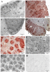
Immunohistochemistry for VEGFA in Embryonic Day (E) 14 testis (magnification, ×200) (A), E14 (×400) (B), E16 (×400) (C), E16 (×200) (D), E19 (×400) (E), P0 (×400) (F), and Postnatal Day 5 (×400) (G). E16 sections with no primary antibody (F) served as negative controls. These results are representative images from 3 different experiments conducted with three to six different sections of tissue from each age group. T = testis; M = mesonephros; c = cord; i = interstitium; S = Sertoli; G = germ.
Figure 3
Figure 3
Immunohistochemistry for KDR (VEGFR2) expression in testes at Embryonic Day (E) 14 (magnification, ×200) (A), E14 (×400) (B), E19 (×400) (C), Postnatal Day (P) 0 (×400) (D), and P5 (×400) (E). E16 controls with no primary antibody (F) served as negative controls. These results are representative images from three different experiments conducted with three to six different sections of testis tissue from each age group. T = testis; M = mesonephros; c = cord; i = interstitium; S = Sertoli; G = germ. Arrows point to Sertoli and germ cells.
Figure 4
Figure 4
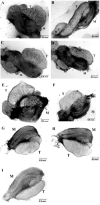
Effect of the VEGFR signal transduction inhibitors VEGFR-TKI, SU1498, and Je-11 on seminiferous cord formation in organ cultures of rat testis obtained on Embryonic Day 13. Organ cultures contained vehicle controls (DMSO) (A, C, E, G) or specimens treated daily with 8 μM of VEGFR-TKI (B, D), 40 μM of SU1498 (F), or 10 μM of Je-11 (VEGFA antagonist) (H, I). T = testis; M = mesonephros.
Figure 5
Figure 5
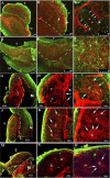
Whole-mount immunohistochemistry of Embryonic Day 13 testis organ culture controls for VEGFR-TKI treatments (A, B, C), testis cultures treated with 8 μM VEGFR-TKI (D-G), vehicle controls for VEGFA antagonist Je-11 (G-L), and testis cultures treated with 10 μM Je-11 (M-O). Cultured organs were labeled with antibodies against laminin (green, basement membrane marker) and PECAM (red, endothelial cell and mitotic germ cell marker). Note that only mitotic germ cells express PECAM; germ cells that are not mitotic do not express PECAM. A representative control organ (A-C), shows both blood vessel patterns (arrows, B) and seminiferous cord formation (arrows, C). Mitotic germ cells can be seen within the seminiferous cords (*, C). Organs treated with 8 μM VEGFR-TKI (D-F) do not have normal vasculature patterns or cord formation. Magnifications are ×100 (A, D), ×200 (B, E), and ×600 (C, F, G). Normal vascular patterns surrounding seminiferous cords within the testis are detected in vehicle controls for Je-11 at magnifications of ×100 (G, J), ×200 (H, K), and ×400 (I, L). At high magnifications (I, K, L), mitotic germ cells (arrows) have organized into cord-like structures and are surrounded by vasculature. The coelomic vessel also expressed PECAM (K, L). In Je-11-treated testes, germ cells are clustered together into smaller, irregular cord patterns with reduced vascular density at magnifications of ×100 (M), ×200 (N), and ×400 (O). T = testis; M = mesonephros; c = cord; CV = coelomic vessel. Dashed lines indicate the boundary between the testis and mesonephros.
Figure 6
Figure 6
Effects of LY 294002 on organ cultures rat testis obtained on Embryonic Day (E) 13. Cultured testes from E13 rats treated with DMSO vehicle control (A, C) developed normal seminiferous cords, whereas testes treated with 10 μM (B) or 15 μM (D) of the PI3K inhibitor LY 294002 developed smaller and occasionally swollen cords (B) or had perturbed cord formation (D). T = testis; M = mesonephros.
Figure 7
Figure 7
Effect of 15 μM of LY 294002 on whole-mount immunohistochemistry on Embryonic Day 13 rat testis and mesonephros organ cultures. A representative control organ (A-C) with germ cells is organized within cords surrounded by vasculature (arrows). The treated testis (D-F) is smaller than the control, and germ cells are not as clearly organized within cords. Vasculature surrounds these cords (F) but is less abundant than that in paired control testes. T = testis; M = mesonephros; c = cords.

References

    1. Jost A, Magre S, Agelopoulou R. Early stages of testicular differentiation in the rat. Hum Genet. 1981;58:59–63. - PubMed
    1. Magre S, Jost A. The initial phases of testicular organogenesis in the rat: an electron microscopy study. Arch Anat Microsc Morphol Exp. 1980;69:297–318. - PubMed
    1. Albrecht KH, Eicher EM. Evidence that Sry is expressed in pre-Sertoli cells and Sertoli and granulosa cells have a common precursor. Dev Biol. 2001;240:92–107. - PubMed
    1. Capel B, Albrecht KH, Washburn LL, Eicher EM. Migration of mesonephric cells into the mammalian gonad depends on Sry. Mech Dev. 1999;84:127–131. - PubMed
    1. McLaren A. Development of the mammalian gonad: the fate of the supporting cell lineage. Bioassays. 1991;13:151–156. - PubMed

Publication types

MeSH terms

Substances

LinkOut - more resources