. 2006 Jun;27(6):558-67.
doi: 10.1002/humu.20337.

The spectrum of WRN mutations in Werner syndrome patients

Affiliations

The spectrum of WRN mutations in Werner syndrome patients

Shurong Huang et al. Hum Mutat. 2006 Jun.

Abstract

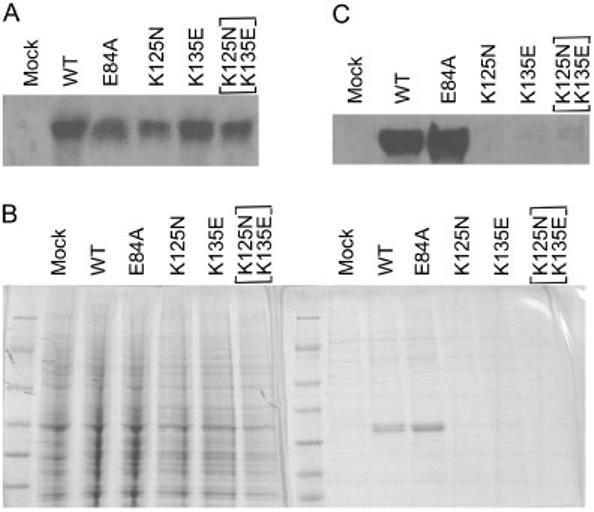
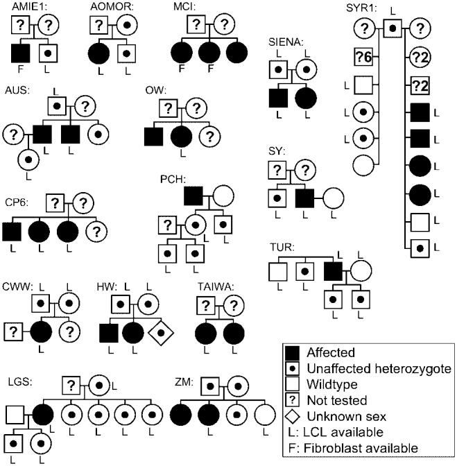
The International Registry of Werner syndrome (www.wernersyndrome.org) has been providing molecular diagnosis of the Werner syndrome (WS) for the past decade. The present communication summarizes, from among 99 WS subjects, the spectrum of 50 distinct mutations discovered by our group and by others since the WRN gene (also called RECQL2 or REQ3) was first cloned in 1996; 25 of these have not previously been published. All WRN mutations reported thus far have resulted in the elimination of the nuclear localization signal at the C-terminus of the protein, precluding functional interactions in the nucleus; thus, all could be classified as null mutations. We now report two new mutations in the N-terminus that result in instability of the WRN protein. Clinical data confirm that the most penetrant phenotype is bilateral ocular cataracts. Other cardinal signs were seen in more than 95% of the cases. The median age of death, previously reported to be in the range of 46-48 years, is 54 years. Lymphoblastoid cell lines (LCLs) have been cryopreserved from the majority of our index cases, including material from nuclear pedigrees. These, as well as inducible and complemented hTERT (catalytic subunit of human telomerase) immortalized skin fibroblast cell lines are available to qualified investigators.


Figures

FIGURE 1
FIGURE 1
Diagram of standard protocols at the International Registry of Werner Syndrome. Typical processing steps of the blood samples are shown.
FIGURE 2
FIGURE 2
Locations of WRN mutations. Exons are designated as boxes 1–35. Introns are indicated by thin lines. The numbers above the exons indicate the nucleotide positions in the WRN cDNA (GenBank accession no. L76937.) with the first ATG being 1. The localizations of the functional domains are indicated. Purple boxes, exonuclease domain; yellow boxes, helicase domain; green boxes, RQC, the RecQ helicase conserved region; pink boxes, HRDC, the Helicase, RnaseD, C-terminal conserved region; and gray box, NLS, the nuclear localization signal; CDS, coding sequence; FS/Ter, frameshift and premature translation termination; IVS, intervening sequence. I-VI indicate the motifs in exonuclease and helicase domains.
FIGURE 3
FIGURE 3
Sequence and structural analysis of the N-terminal region of theWRN protein. A: A multiple sequence alignment of a 3′-5′ exonuclease domain present in the following selected proteins: EcDPOIII, Escherichia coli DNA polymerase III (RCSB code 1J54); EcExoI, E. coli exonuclease I (RCSB 1FXX); EcRNaseT, E. coli RNaseT (RefSeq NP_416169); EcKlenow_1QSL, Klenow fragment of E. coli DNA polymerase I (RCSB 1QSL); HsWRN: Homo sapiensWRN protein (RefSeq NP_000544). Numbers indicate the number of residues not shown explicitly; residues that are conserved in three of the five sequences are highlighted. The annotations below the alignment refer toWRN (HsWRN) and indicate the locations and names of mutations discussed in this study. The annotations above the alignment refer to DNA polymerase I (EcKlenow_1QSL) and refer to features observed in the crystal structure of the Klenow fragment complexed with single-stranded substrate and taken from the 1QSL entry in the PDBsum Resource (www.ebi.ac.uk/thorntonsrv/databases/pdbsum).The arrows and cylinders represent beta-strands and alpha-helices, respectively. Red and blue triangles indicate the locations and names of residues that interact with a metal ion in the active site and DNA substrate respectively. Green triangles mark the residues in the Klenow fragment (K406 and K416) predicted to be equivalent to the WRN mutations K125N and K135E. Yellow triangles mark residues and the boxed region indicates the region shown in the crystal structure. B: Images of the Klenow fragment shown in two different orientations using the same color scheme as in panel A.The thin gray tube is a smoothed representation of the alpha carbon backbone and the small gray spheres denote the N- and C-terminal residues.The N-termini are represented by the gray spheres at the bottoms of the molecules.The single-stranded DNA substrate is shown in cyan (all atom representation).The red and blue side amino acid chains drawn explicitly correspond to the metal and DNA binding residues.The green side chains (K406 and K416) are the positions equivalent to the WRN mutations K125N and K135E; the yellow side chains (E410 and R436) are discussed in the text. C: Images of part of the Klenow fragment shown in two different orientations and corresponding to the region boxed in the alignment. Only atoms in the peptide backbone of Klenow fragment residues 406 to 438 are shown (C: gray, N: blue, O: red).The side chains of four specific residues are drawn explicitly, K406 and K416 (green), and E410 and R436 (yellow).
FIGURE 4
FIGURE 4
K125N and K135E mutations renderWRN proteins unstable. A: Northern blotting of total RNA isolated from insect cells expressing segments of the WRN protein with the indicated mutations. B: Coomassie blue staining of the proteins isolated from the culture. Left panel shows the patterns of 1 mg of total proteins. Right panel shows the patterns of 100 μg of the purified proteins. C:Western analysis of the purified proteins.
FIGURE 5
FIGURE 5
Distribution of the ages of onset of ocular cataracts in molecularly confirmed WS (A), distribution of the ages of the patients at the time of referral to our registry (B), and the distribution of the ages of death (C). X-axes indicate the age in decades, and Y-axes show the number of cases.
FIGURE 6
FIGURE 6
Pedigrees of the WS patients with documented WRN mutations. Family names correspond to those given in Supplementary Table S1.

References

    1. Ahn B, Harrigan JA, Indig FE, Wilson DM, 3rd, Bohr VA. Regulation of WRN helicase activity in human base excision repair. J Biol Chem. 2004;279:53465–53474. - PubMed
    1. Balajee AS, Machwe A, May A, Gray MD, Oshima J, Martin GM, Nehlin JO, Brosh R, Orren DK, Bohr VA. The Werner syndrome protein is involved in RNA polymerase II transcription. Mol Biol Cell. 1999;10:2655–2668. - PMC - PubMed
    1. Bennett RJ, Keck JL. Structure and function of RecQ DNA helicases. Crit Rev Biochem Mol Biol. 2004;39:79–97. - PubMed
    1. Brosh RM, Jr, von Kobbe C, Sommers JA, Karmakar P, Opresko PL, Piotrowski J, Dianova I, Dianov GL, Bohr VA. Werner syndrome protein interacts with human flap endonuclease 1 and stimulates its cleavage activity. EMBO J. 2001;20:5791–5801. - PMC - PubMed
    1. Chen L, Huang S, Lee L, Davalos A, Schiestl RH, Campisi J, Oshima J. WRN, the protein deficient in Werner syndrome, plays a critical structural role in optimizing DNA repair. Aging Cell. 2003a;2:191–199. - PubMed

Publication types