Comparative Study
. 2006 Jul 15;574(Pt 2):583-96.
doi: 10.1113/jphysiol.2006.108308. Epub 2006 May 4.

Transcapillary fluid balance consequences of missing initial lymphatics studied in a mouse model of primary lymphoedema

Affiliations
Comparative Study

Transcapillary fluid balance consequences of missing initial lymphatics studied in a mouse model of primary lymphoedema

Tine V Karlsen et al. J Physiol. .

Abstract

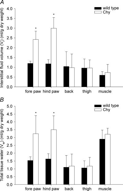
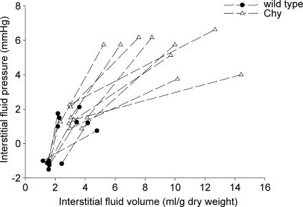
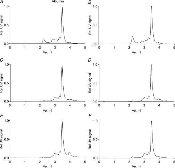
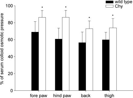
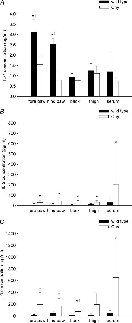
To investigate the phenotypic consequences of a deranged lymphangiogenesis in relation to tissue fluid accumulation and the possible role of inflammation in the pathogenesis of lymphoedema, we measured determinants of transcapillary fluid filtration and inflammatory mediators in the interstitial fluid in genetically engineered Chy mice, a model for primary congenital lymphoedema (Milroy's disease). Although initial lymphatics were not present in dermis in any of the areas studied (fore paw, hind paw, thigh and back skin) interstitial fluid pressure (P(if)), measured with micropipettes, and tissue fluid volumes were significantly increased only in the areas with visible swelling - the fore and hind paw, whereas interstitial colloid osmotic pressure (COP(if)) was increased in all the skin areas examined. A volume load of 15% of body weight resulted in a more pronounced increase in P(if) as well as a four-fold increase in interstitial fluid volume in Chy relative to wild-type (wt) mice, showing the quantitative importance of lymphatics for fluid homeostasis during acute perturbations. A similar level of proinflammatory markers in interstitial fluid in early established lymphoedema (3-4 months) in Chy and wt suggests that inflammation does not have a major pathogenetic role for the development of lymphoedema, whereas a reduced level of the immunomodulatory cytokine interleukin (IL)-4 may result in a reduced immunological defence ability and thus lead to the increase in inflammatory cytokines IL-2 and IL-6 observed at a later stage (11-13 months). Our data suggest that primary lymphoedema results in a high interstitial fluid protein concentration that does not induce an interstitial inflammatory reaction per se, and furthermore shows the paramount importance of the initial lymphatics in tissue fluid homeostasis, especially during perturbations of transcapillary fluid balance.


Figures

Figure 1
Figure 1. Fluid volumes in wt and Chy mice
Interstitial fluid volume (A) and total tissue water (B) in fore paw, hind paw, thigh and back skin together with thigh muscle in Chy and wild type (wt) mice. The values are mean ± s.d. *P < 0.05, significant difference (unpaired t test) between Chy and wt mice within the same area of measurement.
Figure 2
Figure 2. Interstitial fluid pressure in wt and Chy mice
Interstitial fluid pressure in fore paw, hind paw, thigh and back skin together with thigh muscle in Chy and wt mice. The values are mean ± s.d.*P < 0.05, significant difference (unpaired t test) between Chy and wt mice within the same area of measurement.
Figure 3
Figure 3. Response in interstitial fluid pressure to volume loading
Corresponding values for interstitial fluid pressure and volume in hind paw in wt (•) and Chy (▵) mice before and after infusion of saline corresponding to 15% body weight. Values in the same animal have been connected.
Figure 4
Figure 4. Size distribution of macromolecules in serum and interstitial fluid
HPLC chromatograms of the macromolecular distribution pattern in serum and interstitial fluid (IF) from Chy and wt mice. The left panel shows representative chromatograms from wt serum (A), thigh skin IF (C) and hind paw skin IF (E), while the right panel shows Chy serum (B), thigh skin IF (D) and hind paw skin IF (F). The areas under the peaks are normalized as relative to the albumin peak for each chromatogram.
Figure 5
Figure 5. Colloid osmotic pressure in interstitial fluid relative to serum
Colloid osmotic pressure in interstitial fluid isolated from fore paw, hind paw, thigh and back skin in Chy and wt mice. The pressures were calculated as relative to the corresponding serum COP for each mouse. The values are then given as mean ± s.d. *P < 0.05, significant difference (unpaired t test) between Chy and wt mice within the same area of measurement.
Figure 6
Figure 6. Interleukins in serum and interstitial fluid
Interleukins in serum and interstitial fluid from fore paw, hind paw, thigh and back skin of young (3–4 months) and old (11–13 months) adult Chy and wt mice. A, interleukin-4 levels in young adult mice; B, interleukin-2 levels in old adult mice; C, interleukin-6 levels in old adult mice. Values are presented as mean ± s.d. *P < 0.05, significant difference (unpaired t test) between Chy and wt mice within the same area of measurement. †P < 0.05 when compared with corresponding serum.
Figure 7
Figure 7. Initial (LYVE-1)-positive lymphatics in sections from skin
Skin sections (∼5 μm) from Chy and wt mice stained with the lymphatic marker lymphatic vessel endothelial hyaluronan receptor-1 (LYVE-1). The arrows show LYVE-1 staining in the endothelium of lymph vessels. The left panel displays skin sections from wt hind paw (A), fore paw (C), back (E) and thigh (G) while the right panel displays skin sections from Chy hind paw (B), fore paw (D), back (F) and thigh (H). Bar = 50 μm.
Figure 8
Figure 8. Initial (LYVE-1)-positive lymphatics in sections from intact paw
Sections (∼10 μm) from wt (left panel) and Chy (right panel) mice made from intact paw after decalcification, and stained with the lymphatic marker lymphatic vessel endothelial hyaluronan receptor-1 (LYVE-1). The arrows show LYVE-1 staining in the endothelium of lymph vessels. Whereas full-thickness skin in wt mice (A) had lymphatics in the dermis as well as the subcutis, initial lymphatics were located in subcutis only in Chy mice (B). In hindlimb muscle, no difference in staining could be demonstrated between wt (C) and Chy mice (D). Bar = 50 μm.

Comment in

Similar articles

Cited by

References

    1. Adamson RH, Lenz JF, Zhang X, Adamson GN, Weinbaum S, Curry FE. Oncotic pressures opposing filtration across non-fenestrated rat microvessels. J Physiol. 2004;557:889–907. - PMC - PubMed
    1. Alitalo K, Tammela T, Petrova TV. Lymphangiogenesis in development and human disease. Nature. 2005;438:946–953. - PubMed
    1. Aukland K, Reed RK. Interstitial-lymphatic mechanisms in the control of extracellular fluid volume. Physiol Rev. 1993;73:1–78. - PubMed
    1. Baluk P, Tammela T, Ator E, Lyubynska N, Achen MG, Hicklin DJ, Jeltsch M, Petrova TV, Pytowski B, Stacker SA, Yla-Herttuala S, Jackson DG, Alitalo K, McDonald DM. Pathogenesis of persistent lymphatic vessel hyperplasia in chronic airway inflammation. J Clin Invest. 2005;115:247–257. - PMC - PubMed
    1. Bates DO, Levick JR, Mortimer PS. Subcutaneous interstitial fluid pressure and arm volume in lymphoedema. Int J Microcirc Clin Exp. 1992;11:359–373. - PubMed

Publication types

MeSH terms

LinkOut - more resources