. 2006 Sep;20(9):2156-72.
doi: 10.1210/me.2005-0512. Epub 2006 May 4.

Luteinizing hormone-induced RUNX1 regulates the expression of genes in granulosa cells of rat periovulatory follicles

Affiliations

Luteinizing hormone-induced RUNX1 regulates the expression of genes in granulosa cells of rat periovulatory follicles

Misung Jo et al. Mol Endocrinol. 2006 Sep.

Abstract

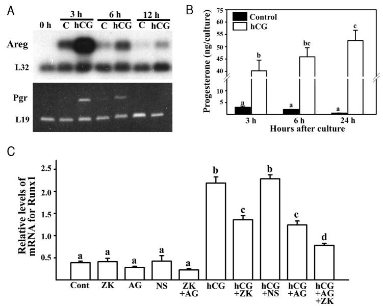
The LH surge induces specific transcription factors that regulate the expression of a myriad of genes in periovulatory follicles to bring about ovulation and luteinization. The present study determined 1) the localization of RUNX1, a nuclear transcription factor, 2) regulation of Runx1 mRNA expression, and 3) its potential function in rat ovaries. Up-regulation of mRNA and protein for RUNX1 is detected in preovulatory follicles after human chorionic gonadotropin (hCG) injection in gonadotropin-treated immature rats as well as after the LH surge in cycling animals by in situ hybridization and immunohistochemical and Western blot analyses. The regulation of Runx1 mRNA expression was investigated in vitro using granulosa cells from rat preovulatory ovaries. Treatments with hCG, forskolin, or phorbol 12 myristate 13-acetate stimulated Runx1 mRNA expression. The effects of hCG were reduced by inhibitors of protein kinase A, MAPK kinase, or p38 kinase, indicating that Runx1 expression is regulated by the LH-initiated activation of these signaling mediators. In addition, hCG-induced Runx1 mRNA expression was inhibited by a progesterone receptor antagonist and an epidermal growth factor receptor tyrosine kinase inhibitor, whereas amphiregulin stimulated Runx1 mRNA expression, demonstrating that the expression is mediated by the activation of the progesterone receptor and epidermal growth factor receptor. Finally, knockdown of Runx1 mRNA by small interfering RNA decreased progesterone secretion and reduced levels of mRNA for Cyp11a1, Hapln1, Mt1a, and Rgc32. The hormonally regulated expression of Runx1 in periovulatory follicles, its involvement in progesterone production, and regulation of preovulatory gene expression suggest important roles of RUNX1 in the periovulatory process.


Figures

Fig. 1
Fig. 1. Expression Profile of RUNX1 in Gonadotropin-Primed Immature Rat Ovaries
A, Immunohistochemical localization for RUNX1 protein during the periovulatory period in gonadotropin-primed immature rat ovaries. Sections of rat ovaries obtained at 0 h (48 h post-PMSG, a and b), 6 h (c, d, i, and j), 12 h (e and f), or 24 h (g and h) after hCG injection were immunostained with the antibody for RUNX1 (AML-1: N-20). The boxed areas in panels a, c, e, g, and i are magnified in panel b, d, f, h, and j, respectively. The tissue section depicted in i and j was incubated with the primary antibody preadsorbed with the blocking peptide for AML1 as a negative control. F, Follicle; Gc, granulosa cell layer; Th, theca cell layer; PF, periovulatory follicles; nCL, newly forming corpus luteum. Arrowheads indicate positive stained nuclei of granulosa cells. Arrows indicate positive stained nuclei of theca cells. Wavy arrows indicate positively stained nuclei of luteal cells. Stars indicate small antral follicles lacking RUNX1 immunoreactivity in the granulosa cells. Original magnification of a, c, e, g, and i is ×150. Original magnification of b, d, f, h, and j panels is ×400. B, Western blot analysis of RUNX1 protein in granulosa cells, residual ovarian tissue, or whole ovaries of immature rats obtained at indicated times after hCG injection. Arrows to the right of panel B) indicate different forms of RUNX1 detected. Each lane was loaded with 25 μl of nuclear fraction (~30 μg) extracted from ovaries of each animal. The membrane was stained with Ponceau-S to show the relative loading of sample protein. The blots (a and b in panel B) are representatives of four separate experiments (n = 4 animals/time point).
Fig. 2
Fig. 2. Expression Profile of RUNX1 in Naturally Cycling Rat Ovaries
A, In situ hybridization analysis of Runx1 mRNA during the periovulatory period in ovaries from naturally cycling rats. Representative bright-field (a, c, and e) and corresponding dark-field (b, d, and f) photomicrographs are depicted. Ovaries were collected at 1600 h (a and b; at the peak of the LH surge) and 2400 h (c and d) on proestrus, and 400 h on estrus (e and f). Arrows in d indicate a small antral follicle expressing Runx1 mRNA in the theca layer. Arrowheads in d indicate Runx1 mRNA expression in periovulatory follicles. Original magnification of all slides is ×40. B, Immunohistochemical localization for RUNX1 during the periovulatory period in ovaries from naturally cycling rats. Sections of rat ovaries obtained at 1600 h (a and b; at the peak of the LH surge) and 2400 h (c and d) on proestrus, and 400 h on estrus (e and f) were immunostained with the RUNX1 antibody (AML-1: N-20). Arrows, arrowheads, and wavy arrows indicate theca cells, granulosa cells, and luteal cells, respectively, that show positive staining for Runx1 protein. Stars indicate small follicles lacking RUNX1 immunoreactivity in the granulosa cells. The boxed areas in panels a, c, and e were magnified in panels b, d, and f, respectively. Original magnification of a, c, and e panels is ×150. Original magnification of b, d, and f panels is ×400. Gc, Granulosa cell layer; Th, theca cell layer; PF, periovulatory follicles; nCL, newly forming corpus luteum; pCL, corpus luteum from previous estrous cycles; F, follicle.
Fig. 3
Fig. 3. Transient Up-Regulation of mRNA and Protein for RUNX1 in Granulosa Cells by hCG in Vitro
A, Autoradiograph of a representative Northern blot analysis shows multiple transcripts for the Runx1 gene and ribosomal protein L32 mRNA in granulosa cells from rat preovulatory ovaries (48 h post-PMSG) cultured in medium alone (Control) or with hCG (1 IU/ml) for 0, 3, 6, 12, or 24 h. The levels of Runx1 mRNA were calculated by combining the intensity of all three different transcripts of the Runx1 gene detected in the Northern blot. Relative levels of Runx1 mRNA were normalized to the L32 band in each sample (mean ± sem; n = 3 independent culture experiments). Bars with no common superscripts are significantly different (P < 0.05). B, Western blot analysis of RUNX1 protein in preovulatory granulosa cells cultured in medium alone (Control) or with hCG for 3, 6, 12, or 24 h. Arrows to the right of panel B indicate the different forms of RUNX1 detected. Protein concentrations were controlled by plating an equal number of cells per well for each treatment and then loading an equal volume (30 μl) of nuclear extracts to each lane. The membrane was stained with Ponceau-S to show the relative loading of sample protein. Panel B is a representative of four separate culture experiments. C, Control.
Fig. 4
Fig. 4. Regulation of Runx1 mRNA Expression by Agonists or Inhibitors of Various Intracellular Signaling Modulators in Granulosa Cells in Vitro
A, Autoradiograph of a representative Northern blot analysis shows mRNA for Runx1 and ribosomal protein L32 in granulosa cells from rat preovulatory ovaries (48 h post-PMSG) cultured in medium alone (Cont) or with hCG (1 IU/ml), forskolin (FSK, 10 μm), PMA (20 nm), or FSK + PMA for 6 h. The levels of Runx1 mRNA were measured as described in Fig. 3. Relative levels of Runx1 mRNA were normalized to the L32 band in each sample (mean ± sem; n = 7 independent culture experiments). B, Granulosa cells from rat preovulatory ovaries (48 h post-PMSG) were cultured with medium alone (Cont), inhibitors of various signaling molecules [an inhibitor of PKA (H89, 10 μm), PKC (GF109203X[GF], 1 μm), phosphatidylinositol 3-kinase (LY294002[LY], 25 μm), MEK (PD98059[PD], 20 μm), and p38 kinase (SB2035850[SB], 20 μm)], hCG, or hCG + inhibitors of various signaling molecules for 6 h. Levels of Runx1 mRNA were measured by Northern blot analyses. Relative levels of Runx1 mRNA were normalized to the L32 band in each sample (mean ± sem; n = 4 independent culture experiments). Bars with no common superscripts are significantly different (P < 0.05). C, The experimental results are summarized in the diagram. The LH surge or an ovulatory dose of hCG stimulates Runx1 mRNA expression via activating PKA, MEK, and p38 kinase. PI3K, Phosphatidylinositol 3-kinase; PLC, phospholipase C.
Fig. 5
Fig. 5. Complete Inhibition of the hCG-Induced Runx1 mRNA Accumulation by Cyclohexamide
Granulosa cells from rat preovulatory ovaries (48 h post-PMSG) were cultured in medium alone (Cont) or with cyclo-hexamide (1 μg/ml, CHX), hCG (1 IU/ml), or hCG + CHX for 6 h. Relative levels of Runx1 mRNA were normalized to the L32 band in each sample in Northern blots (mean ± sem; n = 4). Bars with no common superscripts in each panel are significantly different (P < 0.01).
Fig. 6
Fig. 6. Regulation of Runx1 mRNA Expression by Hormones
A, A representative Northern blot and RT-PCR show a transient increase in levels of mRNA for Areg and Pgr, respectively. L32 and L19 were used as an internal control for each assay. Granulosa cells from rat preovulatory ovaries (48 h post-PMSG) were cultured in medium alone (C) or with hCG for 0, 3, 6, 12, or 24 h. B, hCG stimulates progesterone production in cultured granulosa cells. Concentrations of progesterone were measured in preovulatory granulosa cell conditioned culture media collected at 3, 6, or 24 h. Bars with no common superscripts are significantly different (P < 0.05). C, hCG-induced Runx1 mRNA expression was reduced by a PGR antagonist (ZK98299) and an EGF receptor tyrosine kinase-selective inhibitor (AG1478), as determined by Northern blot analyses. Granulosa cells from rat preovulatory ovaries (48 h post-PMSG) were cultured in medium alone (Cont) or with NS-398 (1 μm; NS, a specific inhibitor of prostaglandin-endoperoxide synthase 2), ZK98299 (1 μm; ZK, a progesterone receptor antagonist), AG1478 (1 μm; AG, an EGF receptor tyrosine kinase-selective inhibitor), ZK + AG, hCG (1 IU/ml), hCG + NS, hCG + ZK, hCG + AG, hCG + ZK + AG for 6 h. Bars with no common superscripts in each panel are significantly different (P < 0.05).
Fig. 7
Fig. 7. Induction of Runx1 mRNA by AREG Was Partially Mediated by the Activation and Induction of PGR
Granulosa cells from rat preovulatory ovaries (48 h post-PMSG) were cultured in medium alone (Cont) or with agonists and/or inhibitors for 6 h to detect Runx1 mRNA and 4 h to measure the levels of Pgr mRNA. Concentrations of agonists and inhibitors are as follows: hCG (1 IU/ml), AREG (0.1 mg/ml), AG1478 (AG1, 1 μm; AG3, 3 μm), ZK98299 (1 μm, ZK), and cyclohexamide (1 μg/ml, CHX). The levels of Runx1 and Pgr mRNA were determined by Northern blot analyses. L32 was used as an internal control. Relative levels of mRNA for the gene were normalized to the L32 band in each sample (mean ± sem; n = 3 or more independent culture experiments). A, The activation of EGF-receptor by AREG stimulated Runx1 mRNA expression, and cyclohexamide blocked AREG-induced Runx1 mRNA expression. B, The stimulatory effect of AREG on Runx1 mRNA expression was inhibited by ZK98299. C, AREG stimulated Pgr mRNA expression. D, The stimulatory effect of hCG on Pgr mRNA expression was reduced by AG1478. Bars with no common superscripts in each panel are significantly different (P < 0.05). E, Hypothetical model of Runx1 mRNA regulation by LH. Cont, Control; EGF-R, EGF receptor.
Fig. 8
Fig. 8. Effects of Reduction in RUNX1 by Runx1 siRNA in Cultured Granulosa Cells
Granulosa cells obtained from rat preovulatory ovaries (48 h post-PMSG) were cultured with hCG (1 IU/ml) + control siRNA (scrambled siRNA) or hCG + Runx1 siRNA. A, Autoradiograph of a representative Northern blot shows mRNA for Runx1 and L32 in the granulosa cells cultured for 4 and 24 h. Relative levels of Runx1 mRNA were normalized to the L32 band in each sample (mean ± sem; n = 7 independent culture experiments). B, Western blot analysis of RUNX1 protein in preovulatory granulosa cells cultured with hCG (1 IU/ml) + control siRNA (scrambled siRNA) or hCG + Runx1 siRNA for 10 h. Arrows to the right of panel B indicate the different forms of Runx1 detected. Protein concentrations were controlled by plating an equal number of cells per well in each treatment and then loading an equal volume (30 μl) of nuclear extract in each lane. The membrane was stained with Ponceau-S to show the relative loading of sample protein. Panel B is a representative of four separate culture experiments. C, Concentrations of progesterone in granulosa cell culture media collected at 24 h after hCG treatment (mean ± sem; n = 6 independent culture experiments). D, Autoradiograph of a representative Northern blot shows mRNA for Cyp11a1, Timp1, and L32 in granulosa cells cultured for 4 and 24 h. Relative levels of Cyp11a1 mRNA were normalized to the L32 band in each sample (mean ± sem; n = 7 independent culture experiments). *, P < 0.001; **, P < 0.01. Cont, Control.
Fig. 9
Fig. 9. Regulation of Periovulatory Gene Expression by Suppression of RUNX1 via siRNA in Cultured Granulosa Cells
Granulosa cells obtained from rat preovulatory ovaries (48 h post-PMSG) were cultured with hCG (1 IU/ml) + control siRNA (scrambled siRNA) or hCG + Runx1 siRNA. A, Autoradiograph of a representative Northern blot shows mRNA for Runx1 and L32 in the granulosa cells cultured with hCG + siRNA for 4, 10, and 24 h. B, Autoradiograph of a representative Northern blot shows mRNA for Runx1, Cdkn1a, Mt1a Hapln1, Rgc32, and L32 (internal control) in the granulosa cells cultured with hCG + siRNA for 10 and 24 h. Relative levels of Mt1a (C), Hapln1 (D), and Rgc32 (E) mRNA were normalized to the L32 band in each sample (mean ± sem; n = 4 independent culture experiments). *, P < 0.001; **, P < 0.01. Cont, Control.

Similar articles

Cited by

References

    1. Espey LL, Richards JS. Temporal and spatial patterns of ovarian gene transcription following an ovulatory dose of gonadotropin in the rat. Biol Reprod. 2002;67:1662–1670. - PubMed
    1. Richards JS, Russell DL, Ochsner S, Espey LL. Ovulation: new dimensions and new regulators of the inflammatory-like response. Annu Rev Physiol. 2002;64:69–92. - PubMed
    1. Richards JS. Ovulation: new factors that prepare the oocyte for fertilization. Mol Cell Endocrinol. 2005;234:75–79. - PubMed
    1. Robker RL, Russell DL, Espey LL, Lydon JP, O’Malley BW, Richards JS. Progesterone-regulated genes in the ovulation process: ADAMTS-1 and cathepsin L proteases. Proc Natl Acad Sci USA. 2000;97:4689–4694. - PMC - PubMed
    1. Sterneck E, Tessarollo L, Johnson PF. An essential role for C/EBPβ in female reproduction. Genes Dev. 1997;11:2153–2162. - PMC - PubMed

Publication types

MeSH terms