. 2006 Sep;20(9):2062-79.
doi: 10.1210/me.2005-0316. Epub 2006 May 4.

Androgens, progestins, and glucocorticoids induce follicle-stimulating hormone beta-subunit gene expression at the level of the gonadotrope

Affiliations

Androgens, progestins, and glucocorticoids induce follicle-stimulating hormone beta-subunit gene expression at the level of the gonadotrope

Varykina G Thackray et al. Mol Endocrinol. 2006 Sep.

Abstract

FSH is produced by the pituitary gonadotrope to regulate gametogenesis. Steroid hormones, including androgens, progestins, and glucocorticoids, have all been shown to stimulate expression of the FSHbeta subunit in primary pituitary cells and rodent models. Understanding the molecular mechanisms of steroid induction of FSHbeta has been difficult due to the heterogeneity of the anterior pituitary. Immortalized LbetaT2 cells are a model of a mature gonadotrope cell and express the endogenous steroid receptor for each of the three hormones. Transient transfection of each receptor, along with ligand treatment, stimulates the mouse FSHbeta promoter, but induction is severely diminished using receptors that lack the ability to bind DNA, indicating that induction is likely through direct DNA binding. All three steroid hormones act within the first 500 bp of the FSHbeta promoter where six putative hormone response elements exist. The -381 site is critical for FSHbeta induction by all three steroid hormones, whereas the -197 and -139 sites contribute to maximal induction. Interestingly, the -273 and -230 sites are also necessary for androgen and progestin induction of FSHbeta, but not for glucocorticoid induction. Additionally, we find that all three receptors bind the endogenous FSHbeta promoter, in vivo, and specifically bind the -381 site in vitro, suggesting that the binding of the receptors to this element is critical for the induction of FSHbeta by these 3-keto steroid hormones. Our data indicate that androgens, glucocorticoids, and progestins act via their receptors to directly activate FSHbeta gene expression in the pituitary gonadotrope.


Figures

Fig. 1
Fig. 1. Androgens, Progestins, and Glucocorticoids Induce FSHβ Gene Expression in Immortalized Gonadotropes
A, RT-PCR was used to detect expression of the steroid receptors in LβT2 cells. Amplified AR is visible at 550 bp, PR at 406 bp, and GR at 298 bp. In reverse transcription of total RNA isolated from LβT2 cells, + indicates the presence of reverse transcriptase, and − indicates no reverse transcriptase. B, ChIP was performed using cross-linked protein/chromatin from LβT2 cells and antibodies directed against AR, PR, and GR or, as a negative control, against nonspecific IgG. Upper panel, PCR primers encompassing the proximal promoter of FSHβ were used to detect precipitation of genomic DNA. Lower panel, PCR primers encompassing the downstream FSHβ coding region were used as a control for specificity. PCR amplification was performed on 0.2% chromatin input (lane 1), and chromatin was precipitated with either mouse IgG (lane 2), AR (lane 3), PR (lane 4), or GR (lane 5) antibodies. C, The −1000FSHβluc reporter gene was transiently transfected into LβT2 cells along with 200 ng of the respective steroid receptor expression vectors. After overnight starvation in serum-free media, the cells were treated with 100 nm testosterone, DHT, progesterone, corticosterone, or 17β-estradiol for 24 h. Luciferase activity was normalized to β-galactosidase activity and set relative to the empty reporter vector. The results represent the mean ± sem of at least three experiments performed in triplicate and are presented as fold induction of hormone treatment relative to the vehicle control (ethanol). For cells transfected with AR, and treated with testosterone or DHT, * indicates significantly different from the vehicle-treated control, using one-way ANOVA followed by Tukey’s post hoc test. For LβT2 cells transiently transfected with PR, GR, ERα, or ERβ and treated for 24 h with progesterone, corticosterone, or 17β-estradiol, respectively, * indicates significantly different from the respective vehicle-treated control using Student’s t test. RT, Reverse transcriptase.
Fig. 2
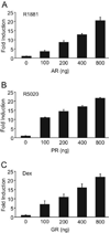
Fig. 2. Androgens, Progestins, and Glucocorticoids Induce Transcription of FSHβ in a Receptor-Dependent Manner
The −1000FSHβluc reporter gene was transiently transfected into LβT2 cells along with increasing receptor concentrations ranging from 0–800 ng/well, as indicated. After overnight starvation in serum-free media, the cells were treated for 24 h with the indicated hormone. The results represent the mean ± sem of at least three experiments performed in triplicate and are presented as fold induction relative to the vehicle control. A, LβT2 cells were transiently transfected with increasing amounts of AR and then treated with 100 nm R1881. B, LβT2 cells were transiently transfected with increasing amounts of PR and then treated with 100 nm R5020. C, LβT2 cells were transiently transfected with increasing amounts of GR and then treated with 100 nm dexamethasone (Dex).
Fig. 3
Fig. 3. Steroid Hormones Mediate Transcription of FSHβ in a Dose-Dependent Manner
The −1000FSHβluc reporter gene was transiently transfected into LβT2 cells along with the respective receptor expression vector indicated on the graph. After overnight starvation in serum-free media, the cells were treated for 24 h with the indicated hormone concentrations. The results represent the mean ± sem of at least three experiments performed in triplicate and are presented as fold induction relative to the vehicle control. A, LβT2 cells were transiently transfected with the AR expression vector and then treated with R1881 concentrations ranging from 10 pm to 100 nm. B, LβT2 cells were transiently transfected with the PR expression vector and then treated for 24 h with 10 pm to 100 nm R5020. C, LβT2 cells were transiently transfected with the GR expression vector and then treated with 100 pm to 1 µm dexamethasone (Dex).
Fig. 4
Fig. 4. Androgens, Progestins, and Glucocorticoids Down-Regulate LHβ Gene Expression
The 1.8LHβluc reporter gene was transiently transfected into LβT2 cells along with the indicated receptor expression vector. After overnight starvation in serum-free media, the cells were treated for 24 h with hormone. The results represent the mean ± sem of at least three experiments performed in triplicate and are presented as fold induction relative to the vehicle control. *, Indicates that the hormone treatment was significantly different from the vehicle-treated control using Student’s t test. A, LβT2 cells were transiently transfected with the AR expression vector and then treated with 100 nm R1881. B, Cells were transiently transfected with the PR expression vector and then treated with 100 nm R5020. C, Cells were transiently transfected with the GR expression vector and then treated with 100 nm dexamethasone (Dex).
Fig. 5
Fig. 5. AR, PR, and GR Require DNA Binding to Facilitate FSHβ Gene Expression
A, C, and E, Wild-type and mutant steroid receptors were overexpressed, and protein levels were analyzed by Western blot to ensure that wild-type and mutant receptors were expressed at a similar level. The arrowhead denotes the band specific for the respective steroid receptors, and the nonspecific bands demonstrate equal protein loading. A, WT AR, mutant AR (ARC562G), or empty vector control (pSG5) were overexpressed in LβT2 cells. B, WT PR, mutant PR (PRC577A) or empty vector control (pCMV5) were overexpressed in Cos-1 cells. C, WT GR, mutant GR (GRdim4) or empty vector control (pSG5) were overexpressed in Cos-1 cells. B, D, and F, −1000FSHβluc reporter gene was transiently transfected into LβT2 cells along with the wild-type or mutant receptor as indicated. After overnight starvation in serum-free media, the cells were treated for 24 h with hormone. The results represent the mean ± sem of at least three experiments performed in triplicate and are presented as fold induction relative to the vehicle control. *, Indicates that the hormone treatment was significantly different from the vehicle-treated control using Student’s t test. B, LβT2 cells were transiently transfected with the wild-type or mutant AR (ARC562G) expression vector and then treated with 100 nm R1881. D, Cells were transiently transfected with wild-type or mutant PR (PRC577A) expression vector and then treated with 100 nm R5020. F, Cells were transiently transfected with wild-type GR or GRdim4 mutant expression vector and then treated with 100 nm dexamethasone (Dex). WT, Wild type.
Fig. 6
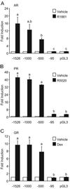
Fig. 6. Induction of FSHβ by Steroid Hormone Receptors Maps to a Region Between −500 and −95 bp of the Proximal Promoter
The −1526FSHβluc, −1000FSHβluc, −500FSHβluc, or −95FSHβluc reporter genes were transiently transfected into LβT2 cells along with the indicated receptor expression vector. After overnight starvation in serum-free media, the cells were treated for 24 h with hormone. The results represent the mean ± sem of at least three experiments performed in triplicate and are presented as fold induction relative to the vehicle control. Significantly different levels of hormone induction among truncations are indicated by differing letters, a, b, or c as determined by one-way ANOVA followed by Tukey’s post hoc test. A, LβT2 cells were transiently transfected with the reporter genes and the AR expression vector after which the cells were treated with 100 nm R1881. B, LβT2 cells were transiently transfected with the reporter genes and the PR expression vector and then the cells were treated with 100 nm R5020. C, LβT2 cells were transiently transfected with the reporter genes and the GR expression vector after which the cells were treated with 100 nm dexamethasone (Dex).
Fig. 7
Fig. 7. Conservation of Putative HREs in the Proximal FSHβ Promoter
Putative HREs from the mouse, rat, ovine, and human FSHβ promoters are aligned 5′ to 3′. The 5′-start of the HRE in each respective species is given on the left. Bold letters indicate homology to the mouse sequence. Boxes highlight conserved G/C residues. References for the studies in which the respective HREs have been characterized are listed on the right.
Fig. 8
Fig. 8. Multiple HREs Play Roles in the Induction of FSHβ by AR, PR, and GR
The wild-type −1000FSHβluc reporter gene, or one of the six mutants, was transiently transfected into LβT2 cells along with the indicated receptor expression vector. After overnight starvation in serum-free media, the cells were treated for 24 h with hormone. The results represent the mean ± sem of at least three experiments performed in triplicate and are presented as fold induction relative to the vehicle control. *, Indicates that the hormone treatment was significantly different from the vehicle-treated control using Student’s t test. #, Indicates that the induction of the mutant reporter gene is significantly different from the induction of the wild-type reporter gene using one-way ANOVA followed by Tukey’s post hoc test. A, LβT2 cells were transiently transfected with the indicated reporter gene and the AR expression vector and then treated with 100 nm R1881. B, LβT2 cells were transiently transfected with the indicated reporter gene and the PR expression vector and then treated with 100 nm R5020.C, LβT2 cells were transiently transfected with the indicated reporter gene and the GR expression vector and then treated with 100 nm dexamethasone (Dex). WT, Wild type.
Fig. 9
Fig. 9. Ligand-Bound AR, PR, and GR All Bind Specifically to the −381 HRE
Whole-cell extracts containing overexpressed AR, PR, or GR from baculovirus-infected insect cells were incubated with the −139, −230, or −381 probe and tested for complex formation in EMSA. The relevant steroid receptor-DNA complex on the −381 element is shown in lane 3 and on the mutated −381 element (mutation as in Fig. 8), whereas the antibody supershift is shown in lane 5, IgG control (lane 6), self-competition (lane 7), mutant competition (lane 8), and competition with a consensus HRE (HRE) (lane 9). A, Probes were incubated with overexpressed AR. B, Probes were incubated with overexpressed PR. C, Probes were incubated with overexpressed GR. Ab, Antibody; Comp, competition.
Fig. 10
Fig. 10. PR Binds the −197 HRE
Whole-cell extracts containing overexpressed PR from baculovirus-infected insect cells were incubated with the −273, −230, −197, or −139 probes and tested for complex formation in EMSA. Antibodies (Ab) are indicated with the left lane containing no antibody (−), the middle lane containing PR antibody (PR), and the right lane containing a nonspecific IgG control antibody (IgG). The antibody supershift is marked with an arrowhead.
Fig. 11
Fig. 11. Specific Protein Complexes from LβT2 Nuclear Extracts Bind the −273, −230, −197, and −139 HREs
Nuclear extracts prepared from LβT2 cells were incubated with the −273, −230, −197, or −139 probes and tested for specific complex formation in EMSA. Unlabeled competitors (500×) (Comp) were added to test the specificity of complex formation as indicated. A, −273 probe with no competition (−), self-competition (273), or mutant competition (273m). B, −230 probe with no competition (−), self-competition (230), or mutant competition (230m). C, −197 probe with no competition (−), self-competition (197), or mutant competition (197m). D, −139 probe with no competition (−), self-competition (139), or mutant competition (139m). Complexes competed by self-competition, but not by mutant competition, are indicated with arrows.

Similar articles

Cited by

References

    1. Kumar TR, Wang Y, Lu N, Matzuk MM. Follicle stimulating hormone is required for ovarian follicle maturation but not male fertility. Nat Genet. 1997;15:201–204. - PubMed
    1. Ma X, Dong Y, Matzuk MM, Kumar TR. Targeted disruption of luteinizing hormone β-subunit leads to hypogonadism, defects in gonadal steroidogenesis, and infertility. Proc Natl Acad Sci USA. 2004;101:17294–17299. - PMC - PubMed
    1. Burns KH, Matzuk MM. Minireview: genetic models for the study of gonadotropin actions. Endocrinology. 2002;143:2823–2835. - PubMed
    1. Pierce JG, Parsons TF. Glycoprotein hormones: structure and function. Annu Rev Biochem. 1981;50:465–495. - PubMed
    1. Kaiser UB, Jakubowiak A, Steinberger A, Chin WW. Differential effects of gonadotropin-releasing hormone (GnRH) pulse frequency on gonadotropin subunit and GnRH receptor messenger ribonucleic acid levels in vitro. Endocrinology. 1997;138:1224–1231. - PubMed

Publication types

MeSH terms