. 2006 May 16;103(20):7637-42.
doi: 10.1073/pnas.0601638103. Epub 2006 May 4.

Crystal structure of the SOCS2-elongin C-elongin B complex defines a prototypical SOCS box ubiquitin ligase

Affiliations

Crystal structure of the SOCS2-elongin C-elongin B complex defines a prototypical SOCS box ubiquitin ligase

Alex N Bullock et al. Proc Natl Acad Sci U S A. .

Abstract

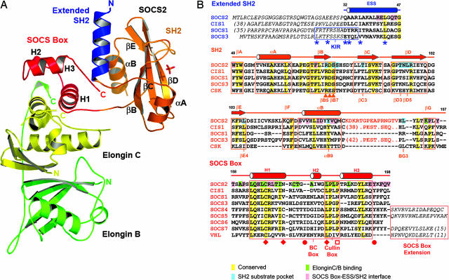
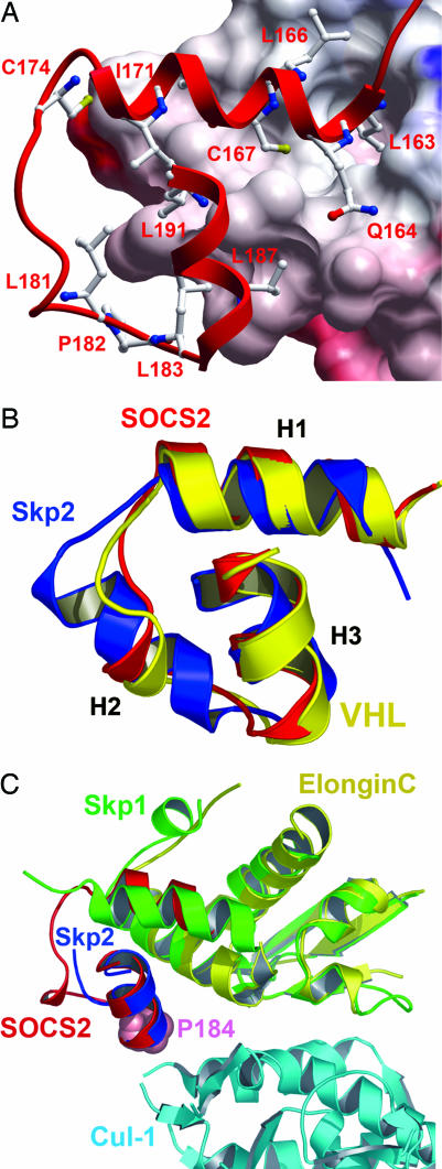
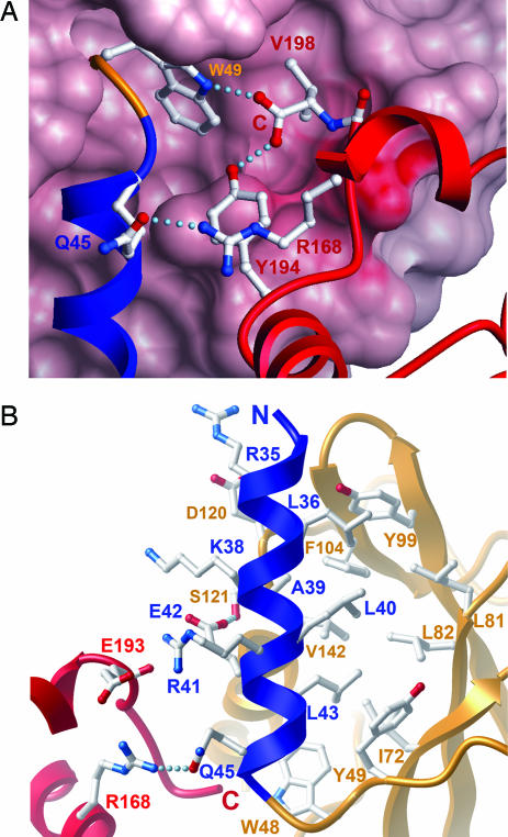
Growth hormone (GH) signaling is tightly controlled by ubiquitination of GH receptors, phosphorylation levels, and accessibility of binding sites for downstream signaling partners. Members of the suppressors of cytokine signaling (SOCS) family function as key regulators at all levels of this pathway, and mouse knockout studies implicate SOCS2 as the primary suppressor. To elucidate the structural basis for SOCS2 function, we determined the 1.9-A crystal structure of the ternary complex of SOCS2 with elongin C and elongin B. The structure defines a prototypical SOCS box ubiquitin ligase with a Src homology 2 (SH2) domain as a substrate recognition motif. Overall, the SOCS box and SH2 domain show a conserved spatial domain arrangement with the BC box and substrate recognition domain of the von Hippel-Lindau (VHL) tumor suppressor protein, suggesting a common mechanism of ubiquitination in these cullin-dependent E3 ligases. The SOCS box binds elongin BC in a similar fashion to the VHL BC box and shows extended structural conservation with the F box of the Skp2 ubiquitin ligase. A previously unrecognized feature of the SOCS box is revealed with the burial of the C terminus, which packs together with the N-terminal extended SH2 subdomain to create a stable interface between the SOCS box and SH2 domain. This domain organization is conserved in SOCS1-3 and CIS1, which share a strictly conserved length of their C termini, but not in SOCS4, 5, and 7, which have extended C termini defining two distinct classes of inter- and intramolecular SOCS box interactions.


Conflict of interest statement

Conflict of interest statement: No conflicts declared.

Figures

Fig. 1.
Fig. 1.
Overall structure of the SOCS2–elongin BC ternary complex and structure-based sequence alignment. (A) The different domains and complex components are differentiated by colors showing the SOCS2 SH2 domain in orange, the ESS in blue, the SOCS box in red, and two elongins in yellow and green. Secondary structure elements discussed in the Results are labeled. (B) Conserved and domain interface residues are highlighted by different colors in the sequence alignment. An unstructured 15-residue insertion in the SOCS2 BG loop is represented by orange text and is the site of predicted PEST sequences in CIS1 and SOCS3 (21). Additional N- and C-terminal extensions are defined by italic text. The positions of mutations that disrupt the SOCS1–JAK2 (22), SOCS2–GHR (11), VHL–elongin C (18), and SOCS1–Cul-5 (16) interactions are indicated by asterisks, triangles, diamonds, and open squares, respectively. Filled circles mark SOCS3 phosphorylation sites, which disrupt the SOCS3–elongin C interaction (20). The SOCS2 SH2 structure is most similar to the structures of C-terminal Src kinase (PDB ID code 1K9A) and Csk-homologous kinase (PDB ID code 1JWO).
Fig. 2.
Fig. 2.
Comparative structural interactions and conservation of the SOCS box, BC box, and F box. (A) SOCS2 binds elongin C (electrostatic surface shown) in a hydrophobic interface of ≈2,200 Å2 (for clarity, elongin B is not shown). Deep pockets accommodate residues from SOCS2 H1 (L163 and C167). (B) The three core helices of the SOCS box (red) show a remarkable structural conservation with the VHL BC box (yellow) and Skp2 F box (blue), forming a common structural motif to link E3 substrate recognition domains with the ubiquitin ligase complex. (C) The crystal structure of the Skp2-Skp1–Cul-1–Rbx1 ternary complex provides a template for the study of SOCS box–elongin C binding to Cul-5 (38). Because of different packing arrangements in their respective complexes, Skp1 shows structural and functional similarity to elongin C and SOCS2 H1, whereas H1 from Skp2 provides equivalence to SOCS2 H3 [consistent with previous Skp2-Skp1 comparison with VHL (39)]. SOCS2 P184 (shown in space-fill) occurs at the likely Cul-5 interface and terminates the “LPXP” cullin box. Mutation of this position in SOCS1 determines Cul-5 interaction (16).
Fig. 3.
Fig. 3.
SOCS2 SH2 domain and binding of GHR phosphopeptide PVPDpYTSIHIV determined by ITC. (A) The SOCS2 substrate pocket has the common hydrophobic cluster at the +3 site, including L95 (βD6), L106 (βE4), Y129 (αB9), and L150 (BG3). The binding site of the phosphotyrosine moiety is indicated by the presence of a bound sulfate ion. Hydrogen bonds are shown as dotted lines. (B) Binding thermodynamic data determined by ITC showed that the GHR-derived phosphopeptide bound with an affinity of 1.6 μM.
Fig. 4.
Fig. 4.
Unpredicted packing arrangements and burial of the SOCS2 N and C termini. (A) The side chain of V198 at the C terminus packs into a deep pocket in the back of the SH2 domain, and the terminal carboxyl forms hydrogen bond interactions with W48 (SH2) and Y194 (SOCS box). This conformation is stabilized by R168, which is strictly conserved within the SOCS family but not within the VHL BC box or Skp2 F box. (B) The ESS forms a single amphipathic helix (blue) that packs between the SH2 (orange) and SOCS box (red) domains with hydrophobic and electrostatic interactions, respectively, that bury a surface area of ≈1,200 Å2.

References

    1. Flores-Morales A., Greenhalgh C. J., Norstedt G., Rico-Bautista E. Mol. Endocrinol. 2005;20:241–253. - PubMed
    1. van Kerkhof P., Smeets M., Strous G. J. Endocrinology. 2002;143:1243–1252. - PubMed
    1. Zhang Y., Jiang J., Black R. A., Baumann G., Frank S. J. Endocrinology. 2000;141:4342–4348. - PubMed
    1. Ram P. A., Waxman D. J. J. Biol. Chem. 1997;272:17694–17702. - PubMed
    1. Metcalf D., Greenhalgh C. J., Viney E., Willson T. A., Starr R., Nicola N. A., Hilton D. J., Alexander W. S. Nature. 2000;405:1069–1073. - PubMed

Publication types

MeSH terms

Associated data