. 2006 May 16;103(20):7694-9.
doi: 10.1073/pnas.0511243103. Epub 2006 May 4.

The bundling activity of vasodilator-stimulated phosphoprotein is required for filopodium formation

Affiliations

The bundling activity of vasodilator-stimulated phosphoprotein is required for filopodium formation

Antje Schirenbeck et al. Proc Natl Acad Sci U S A. .

Abstract

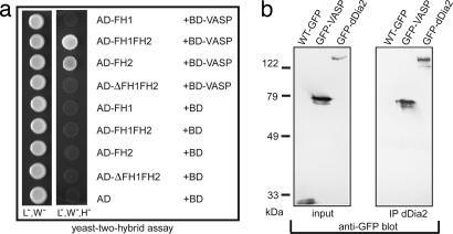
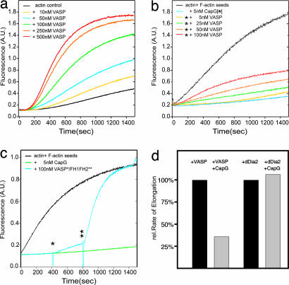
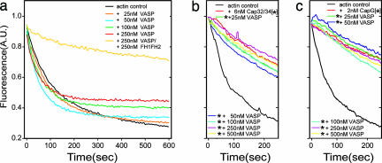
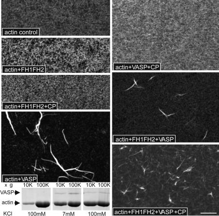
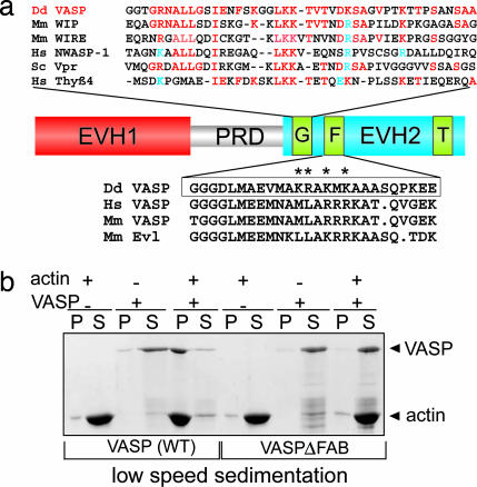
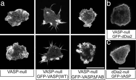
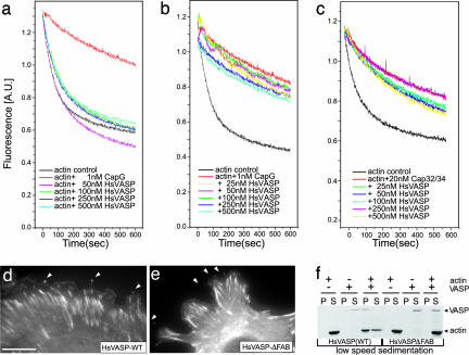
Filopodia are highly dynamic finger-like cell protrusions filled with parallel bundles of actin filaments. Previously we have shown that Diaphanous-related formin dDia2 is involved in the formation of filopodia. Another key player for the formation of filopodia across many species is vasodilator-stimulated phosphoprotein (VASP). It has been proposed that the essential role of VASP for formation of filopodia is its competition with capping proteins for filament barbed-end interaction. To better understand the function of VASP in filopodium formation, we analyzed the in vitro and in vivo properties of Dictyostelium VASP (DdVASP) and extended our findings to human VASP. Recombinant VASP from both species nucleated and bundled actin filaments, but did not compete with capping proteins or block depolymerization from barbed ends. Together with the finding that DdVASP binds to the FH2 domain of dDia2, these data indicate that the crucial role of VASP in filopodium formation is different from uncapping of actin filaments. To identify the activity of DdVASP required in this process, rescue experiments of DdVASP-null cells with mutant DdVASP constructs were performed. Only WT DdVASP, but not a mutant lacking the F-actin bundling activity, could rescue the ability of these cells to form WT-like filopodia. Our data suggest that DdVASP is complexed with dDia2 in filopodial tips and support formin-mediated filament elongation by bundling nascent actin filaments.


Conflict of interest statement

Conflict of interest statement: No conflicts declared.

Figures

Fig. 1.
Fig. 1.
DdVASP interacts with dDia2 in a yeast two-hybrid assay through its FH2 domain. (a) Yeast was transformed with the indicated constructs and tested for interaction by growth on selective media lacking leucine (L), tryptophane (W), or histidine (H) as indicated. AD, activation domain; BD, binding domain. (b) Coimmunoprecipitation of dDia2 and DdVASP. Cell lines expressing GFP, GFP-DdVASP, or GFP-dDia2 (Left) were used for immunoprecipitation with anti-dDia2 polyclonal antibodies. The immunoprecipitates were analyzed in Western blots with anti-GFP antibody mAb 264-449-2.
Fig. 2.
Fig. 2.
DdVASP nucleates actin assembly but does not compete with CP. (a) DdVASP promotes actin polymerization. Actin (1.1 μM) was polymerized in the presence of 0 nM (black line) to 500 nM (red line) DdVASP. (b) DdVASP does not compete with CP. Actin (1.4 μM) was polymerized in the presence of F-actin seeds to avoid de novo nucleation (black line). The presence of 5 nM CP strongly inhibited actin polymerization (light blue line). Addition of increasing amounts of DdVASP to capped filaments showed that DdVASP cannot remove the cap and merely nucleates polymerization of the remaining G-actin. (c) Direct comparison of DdVASP and formin activities. Actin was polymerized in the presence of actin seeds (black line) or capped actin seeds (green and blue lines). After 400 sec (one star) 100 nM DdVASP was added, and after 800 sec (two stars) 100 nM FH1FH2 was added to the same sample. The slope between 400 and 800 sec reflects normal DdVASP-induced de novo nucleation, whereas after addition of FH1FH2 the capped seeds were efficiently uncapped and elongated, leading to a fast increase of F-actin as indicated by the steeper slope. (d) Analysis of nucleating and uncapping activities. The intrinsic nucleation activity of 50 nM DdVASP was measured in the pyrene actin assay, and the slope of fluorescence increase was set to 100% (first bar). After addition of 30 nM CapG the fluorescence increase was strongly inhibited because the newly formed filaments were immediately capped at their barbed ends (second bar). This inhibition was not observed in the presence of FH1FH2 (third and fourth bars) because the formin competes with CP and supports elongation also in the presence of CP.
Fig. 3.
Fig. 3.
DdVASP does not interact with barbed filament ends. (a) Polymerized actin was diluted to 0.1 μM in polymerization buffer alone or in polymerization buffer containing DdVASP or DdVASP and FH1FH2. Note that depolymerization is not attenuated by the addition of DdVASP, indicating that it cannot alter barbed-end kinetics. (b and c) Polymerized actin with free barbed ends or with the barbed ends capped by either Cap32/34 or CapG was diluted to 0.1 μM in polymerization buffer containing 100 mM KCl and the amounts of DdVASP indicated. DdVASP removed neither Cap32/34 nor CapG from the filament ends, because this would have led to fast depolymerization. The stars in b and c indicate the presence of CPs.
Fig. 4.
Fig. 4.
Visualization of F-actin structures formed after incubation with DdVASP, FH1FH2, and CP. G-actin (1.1 μM) was incubated in polymerization buffer in the presence of the indicated proteins (100 nM FH1FH2, 100 nM VASP, and 5 nM CP CapG) on polyl-lysine-coated glass coverslips for 30 min. After fixation the specimens were visualized with tetramethylrhodamine B isothiocyanate–phalloidin. DdVASP induces bundling of F-actin whenever CP is absent or competed off barbed ends by FH1FH2. (Scale bar: 10 μm.) The blot shows a sedimentation assay at 10,000 × g and 100,000 × g to distinguish bundles and actin filaments. Lanes 1 and 2, actin alone plus 100 mM KCl; lanes 3 and 4, actin plus DdVASP at low salt conditions (7 mM KCl); lanes 5 and 6, actin plus DdVASP plus 100 mM KCl. The amount of actin sedimented at 10,000 × g in the presence of DdVASP and 100 mM KCl (lane 5) shows clearly that DdVASP maintains its bundling activity at higher salt concentrations.
Fig. 5.
Fig. 5.
Analysis of Dictyostelium VASP's bundling activity in vitro. (a) The C-terminal EVH2 domain contains three regions: a G-actin binding motif (G), an F-actin binding motif (F), and a region required for tetramerization (T). EVH, Ena/VASP homology; PRD, proline-rich domain. (Upper) Alignment of its G-actin binding site to WH2 (WASP homology domain 2) domains from other G-actin binding proteins: DdVASP, accession no. CAH05068, residues 196–239; Mm WIP, accession no. NP694778, residues 29–70; Mm WIRE, accession no. NP922922, residues 33–75; HsNWASP-1, accession no. NP003932, residues 402–442; Sc Vpr, accession no. NP013441, residues 27–68; Hs Thy-β4, accession no. NP066932, residues 1–41. Identical residues are shown in red, and conserved charged residues are shown in blue. (Lower) The alignment of the F-actin binding regions of VASP from D. discoideum and Ena/VASP-like (Evl) proteins from other species is shown: DdVASP, accession no. CAH05068, residues 264–289; HsVASP, accession no. P50552, residues 297–321; Mm VASP, accession no. P70460, residues 293–317; Mm Evl, accession no. P70429, residues 261–285. The region deleted in the DdVASPΔFAB construct is boxed, and asterisks indicate residues mutated to alanine in the point mutant. (b) Deficiency of F-actin bundling activity by DdVASPΔFAB in a low-speed sedimentation assay. Recombinant WT DdVASP and DdVASPΔFAB (1 μM) were incubated alone or with G-actin (5 μM) in polymerization buffer and spun at 15,000 × g. In contrast to WT DdVASP (left blots), DdVASPΔFAB (right blots) does not cosediment with the F-actin. P, pellet; S, supernatant.
Fig. 6.
Fig. 6.
The F-actin bundling activity of DdVASP is required for filopodium formation in vivo. (a) 3D reconstructions of tetramethylrhodamine B isothiocyanate–phalloidin-labeled knockout and reconstituted cells as indicated. (Scale bar: 5 μm.) Only cells reexpressing WT DdVASP display normal filopodia. (b and c) Cellular distribution of GFP-tagged dDia2 or DdVASP in mutants lacking VASP or dDia2. Confocal sections are shown. (Scale bars: 5 μm.)
Fig. 7.
Fig. 7.
In vitro and in vivo analysis of human VASP. (a) HsVASP does not interact with barbed filament ends. Polymerized pyrene-labeled actin was diluted to 0.1 μM in polymerization buffer alone or in polymerization buffer containing increasing concentrations of HsVASP. Whereas the addition of CapG inhibits depolymerization (red line), even high concentrations of HsVASP do not slow down depolymerization. (b and c) Polymerized actin with either free barbed ends (black lines) or with the barbed ends capped by CapG (b) or Cap32/34 (c) was diluted to 0.1 μM in polymerization buffer containing 100 mM KCl and the amounts of HsVASP indicated. Neither CapG nor Cap32/34 was removed by HsVASP from the filament ends. (d and e) The F-actin binding site is not required for proper cellular localization of vertebrate VASP. Shown are mouse embryonic Mena/VASP double knockout fibroblasts stably expressing either GFP-tagged human WT VASP or a mutant lacking the F-actin binding motif. White arrows mark tips of filopodia. (Scale bar: 20 μm.) (f) Deficiency of F-actin bundling activity by HsVASPΔFAB in a low-speed sedimentation assay. Recombinant WT HsVASP and HsVASPΔFAB (1 μM) were incubated alone or with G-actin (5 μM) in polymerization buffer and spun at 15,000 × g. In contrast to WT HsVASP (left blots), HsVASPΔFAB (right blots) does not cosediment with the F-actin. (P, pellet; S, supernatant). Lanes 1, 2, 7, and 8, actin alone; lanes 3, 4, 9, and 10, VASP alone; lanes 5, 6, 11, and 12, actin plus VASP.

Similar articles

Cited by

References

    1. Zigmond S. H. Curr. Opin. Cell Biol. 2004;16:99–105. - PubMed
    1. Watanabe N., Kato T., Fujita A., Ishizaki T., Narumiya S. Nat. Cell Biol. 1999;1:136–143. - PubMed
    1. Evangelista M., Pruyne D., Amberg D. C., Boone C., Bretscher A. Nat. Cell Biol. 2002;4:260–269. - PubMed
    1. Peng J., Wallar B. J., Flanders A., Swiatek P. J., Alberts A. S. Curr. Biol. 2003;13:534–545. - PubMed
    1. Pellegrin S., Mellor H. Curr. Biol. 2005;15:129–133. - PubMed

Publication types

MeSH terms

Associated data