. 2006 May 5;312(5774):741-4.
doi: 10.1126/science.1125629.

Structure of the multidrug transporter EmrD from Escherichia coli

Affiliations

Structure of the multidrug transporter EmrD from Escherichia coli

Yong Yin et al. Science. .

Erratum in

  • Science. 2007 Sep 21;317(5845):1682

Abstract

EmrD is a multidrug transporter from the Major Facilitator Superfamily that expels amphipathic compounds across the inner membrane of Escherichia coli. Here, we report the x-ray structure of EmrD determined to a resolution of 3.5 angstroms. The structure reveals an interior that is composed mostly of hydrophobic residues, which is consistent with its role transporting amphipathic molecules. Two long loops extend into the inner leaflet side of the cell membrane. This region can serve to recognize and bind substrate directly from the lipid bilayer. We propose that multisubstrate specificity, binding, and transport are facilitated by these loop regions and the internal cavity.


Figures
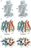
Fig. 1
Fig. 1
Crystallography and structure of EmrD. (A) A portion of the experimental electron density map is shown for H3, H6, and L6-7. The map is contoured to 1 σ. (B) Side view of EmrD. The N- and C-terminus are indicated. (C) View of EmrD looking towards the cytoplasm showing the pseudo two-fold axis relating the N- and C-terminal halves. Transmembrane helices are indicated. The images were created by PyMol.
Fig. 2
Fig. 2
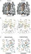
(A) Stereo cut-away view of the hydrophobic internal cavity of EmrD. For clarity, residues 43 to 67 were omitted. Hydrophobicity is shown as a gradation from low (wheat color) to high (brown). Regions that are positive and negative are shown in blue and red, respectively. (B) Inside view of the internal cavity of EmrD characterized by the lining of hydrophobic residues. The N- and C- terminal halves of EmrD and the corresponding residues are colored blue (H1-H6) and orange (H7-H12), respectively. (C) Close view of the selectivity filter region of EmrD. The position of residues that are involved in substrate recognition based on protein sequence homology to other MDR MFS transporters are highlighted (Fig. S1). Residues colored in blue correspond to those in MdfA that when mutated into cysteines either reduce or abolish resistance. Residues in yellow correspond to positions in LmrP that are important for substrate recognition. Val17, shown in red, corresponds to Glu26 in MdfA and Asp23 in FlorR, which are both important for drug recognition. Residues shown in green correspond to cysteine mutations in MdfA that are protected from NEM labeling by substrate. The relative position of the cytoplasm and the internal cavity are indicated.
Fig. 3
Fig. 3
A potential mechanism for hydrophobic substrate transport by EmrD. (A) Drug can enter the internal cavity of the transporter either through the inner membrane leaflet (path ➀) or through the cytoplasm (path ➁). Substrate recognition and binding may be facilitated through the selectivity filter and the internal cavity containing hydrophobic residues. (B) Drug is transported through a rocker-switch alternating access model coupled with H+ antiport. (C) Drug is transported across the lipid bilayer.

References

    1. Putman M, van Veen HW, Konings WN. Microbiol Mol Biol Rev. 2000;64:672. - PMC - PubMed
    1. Naroditskaya V, Schlosser MJ, Fang NY, Lewis K. Biochemical and Biophysical Research Communications. 1993;196:803. - PubMed
    1. Krulwich TA, Quirk PG, Guffanti AA. Microbiological Reviews. 1990;54:52. - PMC - PubMed
    1. Nishino K, Yamaguchi A. J Bacteriol. 2001;183:5803. - PMC - PubMed
    1. Paulsen IT, Brown MH, Skurray RA. Microbiol Rev. 1996;60:575. - PMC - PubMed

Publication types

MeSH terms

Substances

Associated data