. 2006 Jul 15;174(2):213-20.
doi: 10.1164/rccm.200509-1459OC. Epub 2006 May 4.

Haemophilus influenzae forms biofilms on airway epithelia: implications in cystic fibrosis

Affiliations

Haemophilus influenzae forms biofilms on airway epithelia: implications in cystic fibrosis

Timothy D Starner et al. Am J Respir Crit Care Med. .

Abstract

Rationale: Nontypeable Haemophilus influenzae (NTHi) commonly infects patients with cystic fibrosis (CF), especially early in childhood. Bacteria biofilms are increasingly recognized as contributing to bacterial persistence and disease pathogenesis in CF.

Objectives: This study investigated ability of NTHi to form biofilms and its impact on airway epithelia using in vivo and in vitro analyses.

Methods: We evaluated bronchoalveolar lavage fluid from young patients with CF for evidence of NTHi biofilms. To further investigate the pathogenesis of NTHi in respiratory infections, we developed a novel in vitro coculture model of NTHi biofilm formation on polarized human airway epithelial cells grown at the air-liquid interface.

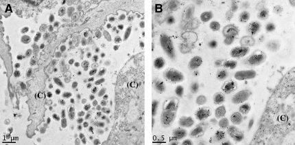
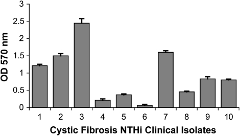
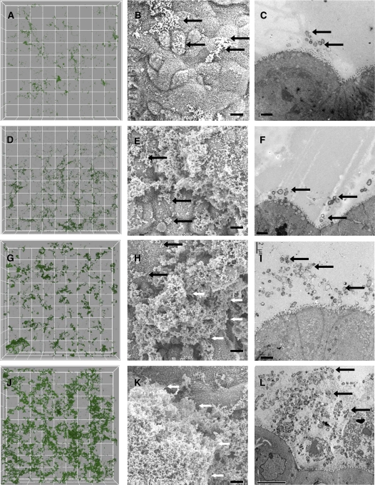
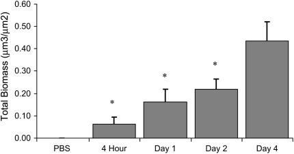
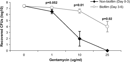
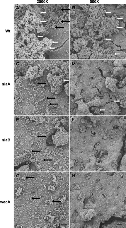
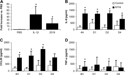
Measurements and main results: In bronchoalveolar lavage fluid samples from young, asymptomatic patients with CF, we found morphologic evidence suggestive of NTHi biofilm formation. In addition, 10 clinical NTHi isolates from patients with CF formed biofilms on plastic surfaces. NTHi formed biofilms on the apical surface of cultured airway epithelia. These biofilms exhibited decreased susceptibility to antibiotics and were adherent to epithelial surfaces. Airway epithelial cells remained viable throughout 4 d of coculture, and responded to NTHi with nuclear factor-kappaB signaling, and increased chemokine and cytokine secretion.

Conclusions: NTHi formed adherent biofilms on the apical surface airway epithelia with decreased susceptibility to antibiotics, and respiratory cells exhibited inflammatory and host defense responses-evidence of a dynamic host-pathogen interaction. The data presented here have implications both for understanding early CF lung disease pathogenesis and for the treatment of early, asymptomatic colonization of patients with CF with H. influenzae.


Figures

<b>Figure 1.</b>
Figure 1.
Evidence for nontypeable Haemophilus influenzae (NTHi) biofilm formation early in cystic fibrosis (CF). Bronchoalveolar lavage fluid (BALF) from an asymptomatic, previously uncolonized 17-mo-old patient with CF revealed bacteria adherent to cell surfaces (CF BALF Patient 3 from Table 1). (A) Immuno–transmission electron microscopy (-TEM) of LR White–embedded CF BALF with a monoclonal antibody against an H. influenzae–specific lipooligosaccharide antigen with small immuno-gold and -silver enhancement, labeled bacteria in the biofilm. (B) Higher power magnification of immunolabeling. “(C)” indicates host cells.
<b>Figure 2.</b>
Figure 2.
Clinical isolates of H. influenzae from patients with CF form biofilms on plastic surfaces. Ten clinical strains of H. influenzae from patients with CF were grown in supplemented brain heart infusion (BHI) broth overnight. Biofilm density was quantified by crystal violet staining as described in Methods, with error bars indicating the standard error of the mean (n = 6). OD = optical density.
<b>Figure 3.</b>
Figure 3.
Airway epithelial cultures infected with NTHi show progressive biofilm formation over time. Samples were inoculated with NTHi and imaged at 4 h, 1 d, 2 d, and 4–5 d. Representative confocal (A, D, G, J), scanning electron microscopic (SEM; B, E, H, K), and TEM images (C, F, I, L) are shown. Four hours (AC): scattered bacteria on the apical surface with virtually no matrix; however, some microcolonies are starting to form; Day 1 (DF): thin matrix production and areas of larger microcolonies; Day 2 (GI): more extensive matrix and larger biofilm structures have formed; Days 4–5 (JL): thick matrix entirely obscuring the apical surface in some areas. Grid boxes on confocal images represent 30 μm2. Scale bars on all SEM images indicate 5 μm. TEM scale bars indicate 2 μm on 4-h–Day 2 images and 10 μm on Days 4–5. Black arrows indicate areas of individual bacteria, and white arrows indicate larger biofilm structures.
<b>Figure 4.</b>
Figure 4.
Quantification of NTHi biofilms in coculture. Quantification of green fluorescent protein–labeled NTHi biofilms grown in coculture on Calu-3 cells as shown in Figure 3. Each datapoint represents the mean biomass of five randomly imaged areas from a single coculture at the times shown. Each graphed value represents the average of the mean biomass values from different coculture experiments, with error bars indicating the standard error of the mean (n = 6–8, except phosphate-buffered saline [PBS], n = 3). *p < 0.001 versus Day 4.
<b>Figure 5.</b>
Figure 5.
NTHi in a biofilm survives high concentrations of gentamicin. NTHi–airway epithelial cocultures were exposed to the indicated concentrations of gentamicin. For the nonbiofilm condition, gentamicin was added from Day 0 at time of NTHi inoculation, until Day 3, when the apical bacteria were recovered. For the biofilm condition, we allowed NTHi to form a biofilm over 3 d in antibiotic-free conditions before exposure to gentamicin on Days 3–6, after which the bacteria were recovered. Mean values for each condition with error bars indicating the standard error of the mean are shown. Significantly more bacteria survived in the biofilm/Day 3–6 biofilm condition compared with the nonbiofilm/Day 0–3 condition at the 10- and 25-mg/ml gentamicin concentrations (n = 3–6).
<b>Figure 6.</b>
Figure 6.
NTHi bacteria deficient in producing a sialylated exopolysaccharide form biofilms poorly. Epithelial cultures were apically infected with NTHi and allowed to form biofilms for 3 d. (A, B) Wild-type NTHi 2019 (Wt) or clinical isolates form robust biofilms. NTHi 2019 with mutations in siaA2019 (siaA; C, D), 2019siaB (siaB; E, F), and 2019wecA (wecA; G, H) had many bacteria on the epithelial surface, but formed biofilms much more poorly. Scale bars on 2,500× and 500× magnifications indicate 5 and 20 μm, respectively. Black arrows indicate areas of individual bacteria, and white arrows indicate larger biofilm structures.
<b>Figure 7.</b>
Figure 7.
Airway epithelial responses to NTHi biofilms. (A) Epithelial cultures were basolaterally transfected with adenovirus containing nuclear factor-κB response elements driving luciferase. Cell lysates from adenovirus transduced cells were obtained 1 d after stimulation with PBS (negative control), 100 ng/ml interleukin (IL)-1β, (positive control), or apical infection with NTHi (n = 11). (BD) ELISA analysis of the basolateral media from control uninfected and NTHi-infected airway epithelial cultures sampled at 4 h, 1 d, 2 d, and 4 d for the indicated chemokines and cytokines (n = 5). Mean values for each condition with error bars indicating the standard error of the mean are shown. *p < 0.05.

Similar articles

Cited by

References

    1. Lam J, Chan R, Lam K, Costerton JW. Production of mucoid microcolonies by Pseudomonas aeruginosa within infected lungs in cystic fibrosis. Infect Immun 1980;28:546–556. - PMC - PubMed
    1. Singh PK, Schaefer AL, Parsek MR, Moninger TO, Welsh MJ, Greenberg EP. Quorum-sensing signals indicate that cystic fibrosis lungs are infected with bacterial biofilms. Nature 2000;407:762–764. - PubMed
    1. Parsek MR, Singh PK. Bacterial biofilms: an emerging link to disease pathogenesis. Annu Rev Microbiol 2003;57:677–701. - PubMed
    1. Nickel JC, Ruseska I, Wright JB, Costerton JW. Tobramycin resistance of Pseudomonas aeruginosa cells growing as a biofilm on urinary catheter material. Antimicrob Agents Chemother 1985;27:619–624. - PMC - PubMed
    1. Jesaitis AJ, Franklin MJ, Berglund D, Sasaki M, Lord CI, Bleazard JB, Duffy JE, Beyenal H, Lewandowski Z. Compromised host defense on Pseudomonas aeruginosa biofilms: characterization of neutrophil and biofilm interactions. J Immunol 2003;171:4329–4339. - PubMed

Publication types