. 2006 Jun 7;25(11):2498-509.
doi: 10.1038/sj.emboj.7601129. Epub 2006 May 4.

Structure of the unusual seryl-tRNA synthetase reveals a distinct zinc-dependent mode of substrate recognition

Affiliations

Structure of the unusual seryl-tRNA synthetase reveals a distinct zinc-dependent mode of substrate recognition

Silvija Bilokapic et al. EMBO J. .

Abstract

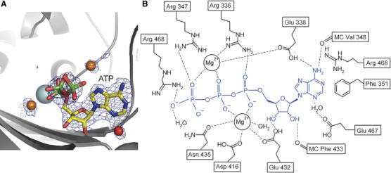
Methanogenic archaea possess unusual seryl-tRNA synthetase (SerRS), evolutionarily distinct from the SerRSs found in other archaea, eucaryotes and bacteria. The two types of SerRSs show only minimal sequence similarity, primarily within class II conserved motifs 1, 2 and 3. Here, we report a 2.5 A resolution crystal structure of the atypical methanogenic Methanosarcina barkeri SerRS and its complexes with ATP, serine and the nonhydrolysable seryl-adenylate analogue 5'-O-(N-serylsulfamoyl)adenosine. The structures reveal two idiosyncratic features of methanogenic SerRSs: a novel N-terminal tRNA-binding domain and an active site zinc ion. The tetra-coordinated Zn2+ ion is bound to three conserved protein ligands (Cys306, Glu355 and Cys461) and binds the amino group of the serine substrate. The absolute requirement of the metal ion for enzymatic activity was confirmed by mutational analysis of the direct zinc ion ligands. This zinc-dependent serine recognition mechanism differs fundamentally from the one employed by the bacterial-type SerRSs. Consequently, SerRS represents the only known aminoacyl-tRNA synthetase system that evolved two distinct mechanisms for the recognition of the same amino-acid substrate.


Figures

Figure 1
Figure 1
Overall structure of SerRS from M. barkeri. (A) Ribbon diagram of the overall crystal structure of aMb-SerRS showing the catalytic core of the two subunits in pink and gray, respectively. The novel RNA-binding domain is in orange with helices proposed to be involved in RNA binding highlighted in red. The noncrystallographic symmetry two-fold axis is in the plane of the paper. (B) Catalytic domain of aMb-SerRS with three class II signature motifs 1, 2 and 3 colored in yellow, blue and green, respectively. Zinc atom and its amino-acid ligands are shown in cyan, as well as the ‘serine ordering loop' (residues 394–410). (C) Topology of the enzyme. Domains and motifs are colored as in (A) and (B). Secondary structure elements are labelled.
Figure 2
Figure 2
Structure-based sequence alignment of aMb-SerRS with selected SerRS sequences from all kingdoms of life and with catalytic domains of closely related ThrRSs. The sequence alignment was firstly generated using the program ClustalX (Thompson et al, 1997) and then manually adjusted based on structural considerations. The sequences are derived from archaea (Mm, Methanococcus maripaludis; Mt, Methanothermobacter thermoautotrophicus; Mk, Methanopyrus kandleri; Mb, Methanosarcina barkeri; Mbu, Methanococcoides burtonii), bacteria (Tt, Thermus thermophilus; Ec, Escherichia coli) and eucarya (Sc, Saccharomyces cerevisiae; Hs, Homo sapiens). One mitochondrial sequence (Bt, Bos taurus) was also included. Amino acids that are completely conserved are in red, whereas those with 80 and 60% conservation are in orange and yellow, respectively. Secondary structural elements are indicated above the alignment with red cylinders for helices and blue arrows for β-sheets. Residues important for zinc ion coordination in aMb-SerRS are marked with asterisks. A green arrow indicates mitochondrial signal sequence. Conserved class II motifs 1, 2 and 3, as well as methanogenic type specific HTH motif, are labeled.
Figure 3
Figure 3
ATP binding by SerRS. (A) Simulated annealing omit FoFc electron density map (resolution 2.0 Å, contour level 2.7σ) together with the refined model: ATP, magnesium ions and surrounding atoms within a sphere of 3 Å were omitted from the model during map calculation. The protein is shown as a ribbon in gray, zinc ion is in cyan, bound magnesium ions and water molecules are shown as orange and red spheres, respectively. The bound ligand is shown in a ball-and-stick representation. (B) Schematic representation of the interactions between the enzyme, ATP and magnesium.
Figure 4
Figure 4
Active site of aMb-SerRS occupied with (A) serine (resolution 2.7 Å, contour level 2.1σ) and (B) seryl-adenylate analogue Ser-AMS (resolution 2.5 Å, contour level 2.2σ). Simulated annealed omit FoFc map calculation as for Figure 3A, orientation and colors are chosen as in Figure 3. Magenta main and side chains undergo serine-induced conformational changes. (C) Schematic representation of the interactions between the enzyme and Ser-AMP. (D) The superposition of Ser-AMS, ATP and serine bound to the active site of aMb-SerRS. ATP is colored in gray, serine in pink and Ser-AMS in an atom-type coloring scheme.
Figure 5
Figure 5
Intersubunit interactions and the role of the HTH motif. (A) A view along the two-fold axis with a dimer in ribbon representation. Additionally, transparent surface representation is shown for one monomer. The view highlights the cross-subunit contacts mediated by the HTH fold. The monomers are shown in pink and gray. (B) Stereoview of interacting residues between HTH fold from one monomer and the N-terminal domain from the symmetry related subunit. The same color code is used as in (A). (C) The superposition of the two subunits, in gray and magenta, by means of the catalytic domain. The figure demonstrates the hinge movement of the tRNA-binding domain and its different orientation with respect to the catalytic core. The view looking down the rotation axis shows that orientation of this domain in two monomers differs by a rotation of ∼20°.
Figure 6
Figure 6
Docking model of tRNA onto Mb-SerRS. (A) Comparison of the overall fold and domain arrangement in the two SerRS representatives: single subunit of atypical (aMb-SerRS in pink) and bacterial (Ec-SerRS in blue) SerRS. Structurally homologous catalytic domains of both SerRSs are in the same orientation. (B) Proposed model of tRNA binding. Two SerRS representatives are shown as dimers and in the same color code as in (A). tRNASer is colored orange and tRNATyr green. The position of the tRNATyr is obtained after superimposition on the core of tRNASer. The figure shows that a hinge movement, as indicated by an arrow, of the M. barkeri N-terminal domain would be required to reach the long variable arm of the tRNASer. (C) Solvent accessible surface representation of the aMb-SerRS dimer colored by electrostatic potential (red for negative and blue for positive) suggests regions involved in the interactions with the negatively charged phosphate backbone of the tRNA molecule.

Similar articles

Cited by

References

    1. Ahel D, Slade D, Mocibob M, Söll D, Weygand-Durasevic I (2005) Selective inhibition of divergent seryl-tRNA synthetases by serine analogues. FEBS Lett 579: 4344–4348 - PubMed
    1. Arnez JG, Dock-Bregeon AC, Moras D (1999) Glycyl-tRNA synthetase uses a negatively charged pit for specific recognition and activation of glycine. J Mol Biol 286: 1449–1459 - PubMed
    1. Baldwin AN, Berg P (1966) Transfer ribonucleic acid-induced hydrolysis of valyladenylate bound to isoleucyl ribonucleic acid synthetase. J Biol Chem 241: 839–845 - PubMed
    1. Baron B, Bock B (1995) The selenocysteine-inserting tRNA species: structure and function. In tRNA: Structure, Biosynthesis and Function, Söll D, RajBhandary U (eds), pp 529–544. Washington, DC: American Society for Microbiology
    1. Beebe K, Merriman E, Ribas de Pouplana L, Schimmel P (2004) A domain for editing by an archaebacterial tRNA synthetase. Proc Natl Acad Sci USA 101: 5958–5963 - PMC - PubMed

Publication types

MeSH terms