. 2006 Jun 7;25(11):2432-42.
doi: 10.1038/sj.emboj.7601122. Epub 2006 May 4.

PIASxbeta acts as an activator of Hoxb1 and is antagonized by Krox20 during hindbrain segmentation

Affiliations

PIASxbeta acts as an activator of Hoxb1 and is antagonized by Krox20 during hindbrain segmentation

Mario Garcia-Dominguez et al. EMBO J. .

Abstract

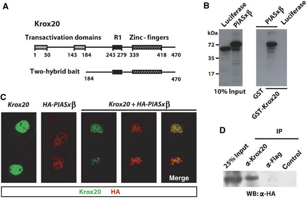
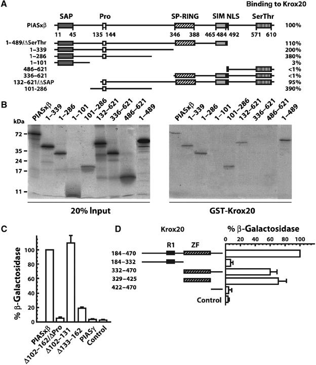
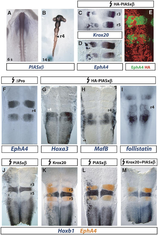
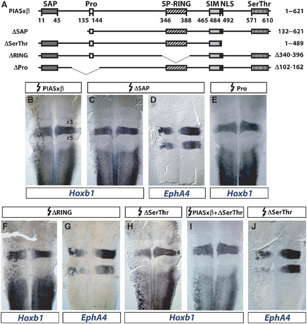
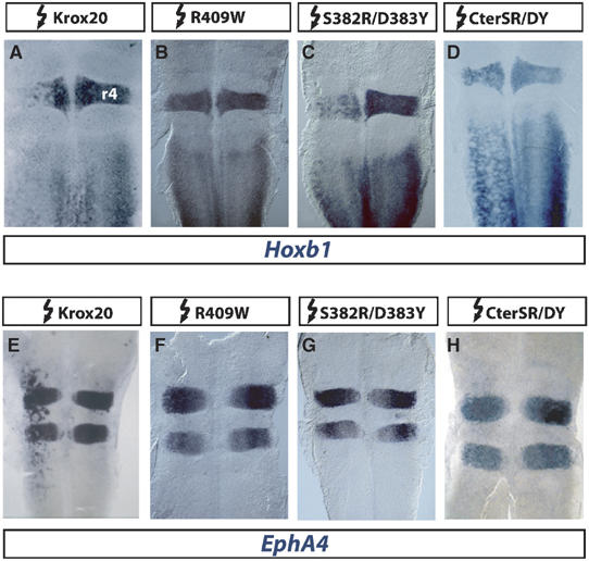
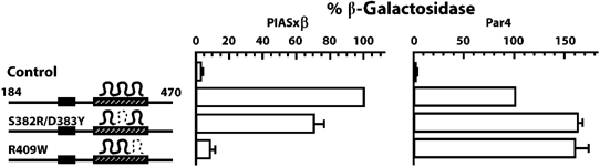
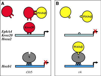
The zinc-finger transcription factor Krox20 constitutes a key regulator of hindbrain development, essential for the formation and specification of rhombomeres (r) 3 and 5. It is in particular responsible for the respective activation and repression of odd- and even-numbered rhombomere-specific genes, which include Hox genes. In this study, we have identified PIASxbeta as a novel direct interactor of Krox20. In addition, we found that PIASxbeta is able to activate the r4-specific gene Hoxb1. Binding of Krox20 prevents this activation, providing a molecular basis for the repression of Hoxb1 by Krox20. The same domain in the Krox20 protein, the zinc-fingers, is involved in DNA binding for transcriptional activation and in interaction with PIASxbeta for transcriptional repression, although the actual precise contacts are different. Our findings add an additional level in the complexity of Hox gene regulation and provide an example of how a single regulator can coordinate the activation and repression of a set of genes by very different mechanisms, acting as a molecular switch to specify cell identity and fate.


Figures

Figure 1
Figure 1
Krox20 interacts with PIASxβ. (A) Schematic structure of the Krox20 protein showing the location of the transactivation domains, NAB interaction domain (R1) and DNA-binding domain (zinc-fingers). Numbers below the line indicate amino-acid positions. The 287 C-terminal amino acids were used as bait in the two-hybrid screening. (B) In vitro binding of Krox20 and PIASxβ. In vitro-translated, 35S-labelled luciferase and PIASxβ were resolved by SDS–PAGE and detected by fluorography. Left: 10% of the input was directly deposited on gel; right: luciferase or PIASxβ retained on GST or GST-Krox20 beads was analysed. PIASxβ specifically binds to GST-Krox20. (C) Colocalization of Krox20 and PIASxβ in the cell nucleus. COS7 cells were transfected with expression vectors encoding Krox20 and/or HA-PIASxβ and 36 h later the proteins were revealed by immunofluorescence analysis (Krox20 is labelled with FITC (green) and HA-PIASxβ with Cy3 (red)). When transfected alone, Krox20 appeared homogenously distributed within the nucleus, whereas Piasxβ localized to nuclear bodies. Cotransfection led to a re-distribution of Krox20, with colocalization of both proteins. (D) Co-immunoprecipitation of Krox20 and PIASxβ. COS7 cells were cotransfected with expression vectors encoding Krox20 and HA-PIASxβ. Cell lysates were subjected to immunoprecipitation with antibodies directed against Krox20 or Flag, or with no antibody (control) and the precipitates were subsequently analysed by Western blotting using an anti-HA antibody.
Figure 2
Figure 2
Identification of the domains involved in PIASxβ/Krox20 interaction. (A) Schematic representation of the PIASxβ structural domains and of the deletion mutants. PIASxβ contains SAP, proline-rich (Pro), SP-RING, SIM domains, an NLS and a C-terminal serine/threonine-rich (Ser/Thr) domain. Numbers below the line indicate amino-acid positions. The amino-acid positions of the extremities of truncated PIASxβ proteins are indicated on the left. The efficiency of the binding to Krox20, indicated in percentage on the right, was estimated from the recovery of each deleted protein after GST-Krox20 pull-down assay (see part B), normalized with wild-type PIASxβ. The PIASxβ interacting region was localized between amino acids 132 and 286. (B) GST-Krox20 pull-down assay performed on deleted PIASxβ. In vitro-translated, 35S-labelled PIASxβ truncated proteins were resolved by SDS–PAGE and detected by fluorography. Left: deposit of 20% of the input proteins; right: PIASxβ proteins recovered after binding to immobilized GST-Krox20 protein. (C) Quantitative yeast two-hybrid assay performed on PIASxβ deletions. PIASγ and a series of PIASxβ deletions fused to the GAL4 activation domain were evaluated for their capacity to bind to a Krox20-Gal4 DNA-binding domain bait by measuring the expression of a lacZ reporter. The β-galactosidase activity was normalized by the level obtained with the wild-type PIASxβ construct. The PIASxβ proline-rich domain appears necessary for the interaction with Krox20. Control corresponds to no PIASxβ insert. (D) Identification of the Krox20 domain required for the interaction with PIASxβ, using the quantitative yeast two-hybrid assay. In this case, a series of Krox20 deletions fused to the Gal4 DNA-binding domain were confronted to the wild-type PIASxβ-Gal4 activation domain fusion and the interaction was recorded by measuring the expression of the lacZ reporter gene. The β-galactosidase activity was normalized by the level obtained with the 184–470 Krox20 deletion construct. The region of interaction with PIASxβ was localized within the zinc-finger domain. Control corresponds to no Krox20 insert.
Figure 3
Figure 3
Ectopic PIASxβ expression antagonizes Krox20 activity and leads to Hoxb1 activation. (A, B) Analysis of PIASxβ expression in chick embryos. Whole-mount in situ hybridization on 6 ss (A) or 14 ss (B) embryos. At 6 ss, PIASxβ is expressed in the neural tube in a decreasing AP gradient. At 14 ss, higher levels of expression are maintained in the anterior CNS up to r2 and in r4. (C–M) Flat-mounted hindbrains from chick embryos electroporated between stages 3 and 8 ss with expression plasmids for the proteins indicated above. The embryos were collected 24 h after electroporation and expression of the markers indicated below was analysed by in situ hybridization (C, D, F–M) or immunochemistry (E). (C, D) Krox20 and EphA4 are repressed in r3 and r5. (E) Double immunohistochemistry with anti-EphA4 (green) and anti-HA (red) antibodies. EphA4-negative patches in r3 and r5 are positive for HA-PIASxβ. Only the left (electroporated) side of the embryo is shown. (F) ΔPro, which has lost the ability to interact with Krox20, does not repress EphA4. (G–I) Hoxa3, MafB and follistatin are not affected by PIASxβ. (J–M) Hoxb1 is ectopically activated by PIASxβ and this activity is antagonized by co-electroporated Krox20 (M). In r3 and r5, Hoxb1 expression domains coincide with the EphA4-negative patches (L). Electroporations were always performed on the left side.
Figure 4
Figure 4
Identification of PIASxβ domains required for Hoxb1 activation. (A) Schematic representation of PIASxβ and deletion mutants (see Figure 2 for details). (B–J) Flat-mounted hindbrains from chick embryos electroporated on the left side with the constructs indicated above and in situ hybridized with the probes indicated below. Whereas deletion of the SP-RING domain does not affect the activity of PIASxβ (B, F, G), elimination of the SAP domain prevents activation of Hoxb1 without affecting the repression of EphA4 (C, D), and deletion of the proline-rich domain prevents activation of Hoxb1 (E). Deletion of the serine/threonine-rich domain does not affect EphA4 repression (J), but transforms PIASxβ into a repressor of Hoxb1 (H), able to antagonize the wild-type protein (I).
Figure 5
Figure 5
Hoxb1 repression by Krox20 does not require DNA binding. (A–H) Flat-mounted hindbrains from chick embryos electroporated on the left side with wild-type and mutant Krox20 constructs as indicated above and in situ hybridized with Hoxb1 or EphA4 probes. (A–D) Whereas the R409W mutation prevents repression of Hoxb1, S382R/D383Y and CterSR/DY conserve this activity, despite their loss of DNA binding. (E–H) In the three mutants, loss of DNA binding correlates with the inability to activate EphA4.
Figure 6
Figure 6
The ability of Krox20 to repress Hoxb1 correlates with its capacity to interact with PIASxβ. The 184–470 Krox20 deletions, in their wild-type version or carrying mutations in the DNA-binding domain, fused to the Gal4 DNA-binding domain were confronted to the Gal4 activation domain fused to PIASxβ or Par4 in the yeast two-hybrid system. The interaction was recorded by measuring the expression of the lacZ reporter gene. The β-galactosidase activities were normalized with the levels obtained with the wild-type construct. Mutant S382R/D383Y, which has retained Hoxb1 repression activity, has also maintained its capacity to interact with PIASxβ, whereas R409W has lost both activities. As a control, interaction with Par4 is not seriously affected in either mutant. Control corresponds to no Krox20 insert.
Figure 7
Figure 7
Schematic representation of the possible crossregulatory interactions between Krox20 and PIASxβ. The relative levels of the two proteins determine the expression of r3/r5- and r4-specific genes (see the Discussion section for details of the model). The circles represent the different proteins and the rectangle the cis-acting elements. (A) Situation in r3/r5. (B) Situation in r4. The interaction of Piasxβ with the Hoxb1 locus may be direct or indirect, via a putative protein (X). The domain of PIASxβ involved in this latter interaction has not been characterized. It is arbitrarily represented as the domain overlapping with the Krox20-binding domain. This representation does not take into account the possibility that PIASxβ might be acting by sequestering a repressor of Hoxb1.

References

    1. Aravind L, Koonin EV (2000) SAP—a putative DNA-binding motif involved in chromosomal organization. Trends Biochem Sci 25: 112–114 - PubMed
    1. Barrow JR, Stadler HS, Capecchi MR (2000) Roles of Hoxa1 and Hoxa2 in patterning the early hindbrain of the mouse. Development 127: 933–944 - PubMed
    1. Birgbauer E, Sechrist J, Bronner-Fraser M, Fraser S (1995) Rhombomeric origin and rostrocaudal reassortment of neural crest cells revealed by intravital microscopy. Development 121: 935–945 - PubMed
    1. Cheng YC, Amoyel M, Qiu X, Jiang YJ, Xu Q, Wilkinson DG (2004) Notch activation regulates the segregation and differentiation of rhombomere boundary cells in the zebrafish hindbrain. Dev Cell 6: 539–550 - PubMed
    1. Choe SK, Sagerstrom CG (2004) Paralog group 1 hox genes regulate rhombomere 5/6 expression of vhnf1, a repressor of rostral hindbrain fates, in a meis-dependent manner. Dev Biol 271: 350–361 - PubMed

Publication types

MeSH terms