. 2006 May;60(4):1026-43.
doi: 10.1111/j.1365-2958.2006.05156.x.

Analysis of FimX, a phosphodiesterase that governs twitching motility in Pseudomonas aeruginosa

Affiliations

Analysis of FimX, a phosphodiesterase that governs twitching motility in Pseudomonas aeruginosa

Barbara I Kazmierczak et al. Mol Microbiol. 2006 May.

Abstract

Type IV pili (Tfp) are polar surface structures of Pseudomonas aeruginosa required for twitching motility, biofilm formation and adherence. One protein required for the assembly of tfp is FimX, which possesses both GGDEF and EAL domains characteristic of diguanylate cyclases and phosphodiesterases respectively. In this work we demonstrate that FimX has phosphodiesterase activity towards bis-(3'-5')-cyclic dimeric guanosine monophosphate (c-di-GMP), but does not show diguanylate cyclase activity. Instead, the imperfect GGDEF domain of FimX likely serves to activate phosphodiesterase activity when bound to GTP, as has recently been described for the Caulobacter crescentus composite GGDEF-EAL protein, CC3396. Bacteria expressing FimX in which either the GGDEF or EAL domain is deleted or mutated have phenotypes indistinguishable from a DeltafimX strain, demonstrating the importance of both domains to function. Previous work has shown that FimX localizes to the bacterial pole. In this work we show that restriction of FimX to a single pole requires intact GGDEF and EAL domains. Deletion of the amino-terminal REC domain of FimX, which contains a putative polar localization signal, results in a protein that still supports intermediate levels of pilus assembly and function. RFP-FimXDeltaREC, unlike RFP-FimX, is no longer localized to the bacterial pole, while transmission electron microscopy shows that surface pili can originate from non-polar sites in this mutant. Although DeltafimX mutants show limited in vitro cytotoxicity, they are as virulent as the wild-type strain in a murine model of acute pneumonia.


Figures

Fig. 1
Fig. 1
FimX is required for normal colony morphology and twitching motility. PA103 and ΔfimX strains were streaked to LB agar plates supplemented with carbenicillin (200 μg m−1). Colonies were photographed under brightfield illumination at 40μ magnification after incubation at 37°C overnight followed by 24 h incubation at room temperature.
Fig. 2
Fig. 2. Construction of FimX domain deletion mutants
A. Schematic showing domains of FimX. Amino acids deleted in each construct (inclusive) or mutated are indicated. The grey bar represents the amino-terminal BB2 epitope tag present in each construct. B. Western blot of whole-cell lysates prepared from ΔfimX strains carrying indicated FimX constructs. Samples were normalized to total protein, separated by SDS-PAGE and transferred to PVDF membranes prior to incubation with anti-BB2 mAb. Calculated molecular weights for each construct are given in parentheses. Migration of molecular weight markers is indicated at left of gel.
Fig. 3
Fig. 3
Total surface pili as determined by ELISA. Bacterial strains were harvested by scraping from LB agar plates after growth overnight at 37°C and resuspended in MEM-lite. Bacterial suspensions were normalized by OD600, then serially diluted. Wells were coated in triplicate with diluted bacteria, fixed, and incubated with primary and secondary antibodies as described in Experimental procedures. Bars indicate mean ± SD of samples diluted to OD600 = 0.003 and are representative of four independent assays.
Fig. 4
Fig. 4
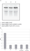
A. Expression of FimX point mutants. Whole-cell lysates were prepared from indicated bacterial strains. Samples were normalized to total protein prior to separation by SDS-PAGE, transferred to PVDF membrane and incubated with anti-BB2 mAb as detailed in Experimental procedures. B. Expression of surface pili as measured by ELISA. Strains were harvested by scraping from agar plates, resuspended in MEM-lite, normalized to OD600, and serially diluted. Wells were coated in triplicate, fixed, and incubated with primary and secondary antibodies as detailed in Experimental procedures. Bars indicate mean ± SD of samples diluted to OD600 = 0.003 and are representative of three independent assays.
Fig. 5
Fig. 5. FimX exhibits phosphodiesterase activity which is stimulated by GTP, but has no detectable DGC activity
A. TLC of DGC assay products. Purified hexahistidine-tagged proteins were assayed in the presence of 10 mM MgCl2 or MnCl2 (as indicated) for the ability to synthesize c-di-GMP from GTP. Reactions were stopped after 30 min by the addition of 0.5 M EDTA and assayed by TLC on PEI-cellulose as detailed in Experimental procedures. B. TLC of phosphodiesterase assay products. Purified hexahistidine-tagged proteins were assayed in the presence of 10 mM MgCl2 or MnCl2 (as indicated), 100 μM GTP (as indicated) and [32P]-c-di-GMP for 30 min. Reactions were stopped by the addition of 0.5 M EDTA and assayed by TLC on PEI-cellulose as detailed in Experimental procedures. No product with Rf(pGpG) was detected when substrate was incubated with MgCl2 and GTP in the absence of protein. C. Phosphodiesterase assay of wild-type FimX and point mutants. Hexahistidine-tagged proteins were incubated with [32P]-c-di-GMP, 10 mM MgCl2, MnCl2, or CaCl2 (as indicated) and 100 μM GTP (as indicated) for 60 min at 25°C. Reaction products were separated by TLC on PEI-cellulose as above. Lanes A–E correspond to: no protein (A); CC3396 (B); FimX (C); FimX(GDSIF→AASIF) (D); FimX(EVL→AAA) (E).
Fig. 6
Fig. 6
Localization of RFP–FimX fusion proteins by indirect immunofluorescence. ΔfimX (panel A) or PA103 (panel B) bacterial colonies expressing indicated RFP–FimX fusion constructs were picked from 2-day-old LB agar plates, resuspended in a drop of ddH2O and spotted onto slides coated with a thin cushion of 1% Gel-Gro gellan. Bacteria were visualized and photographed as detailed in Experimental procedures.
Fig. 7
Fig. 7
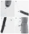
Visualization of tfp by electron microscopy. PA103 pUCP (panels A and B) and ΔfimX pFimXΔREC (panels C and D) bacteria were grown in LB supplemented with carbenicillin to OD600 = 0.6, then gently diluted 20- to 50-fold in ddH2O prior to preparation and staining of grids, as detailed in Experimental procedures. Images were obtained with a Technai 12 Biotwin at magnifications between 13 000x and 30 000x. Negatives were scanned at 1200 dpi, then cropped, assembled and labelled using Photoshop 7.0. Arrows indicate locations at which pili emerge from the cell body.
Fig. 8
Fig. 8. FimX is required for cytotoxicity towards HeLa cells, but not for virulence in the murine model of acute pneumonia
A. LDH release measured from infected HeLa cells. Triplicate wells of HeLa cells were infected with indicated bacterial strains at a moi of 10. Culture supernatants were collected at 2 and 4 hpi and assayed for LDH activity. Bars indicate mean ± SD of a representative experiment. B. Bacterial recovery from infected mice in an acute pneumonia model. Eight- to 10-week-old female C57Bl/6 mice were infected with c. 5 × 10⁵ cfu of PA103 (n = 8) or ΔfimX (n = 10). Mice were euthanized at 16 hpi and the number of bacteria present in lungs and liver were determined as described in Experimental procedures. Results are expressed as the ratio of cfu recovered per g tissue (output) to cfu present in the inoculum (input); each animal is represented by a data point, while the bar shows the geometric mean for each group. The Mann–Whitney test was used to calculate P-values (two-tailed) for each pairwise comparison indicated.

Similar articles

Cited by

References

    1. Aldridge P, Jenal U. Cell cycle-dependent degradation of a flagellar motor component requires a novel-type response regulator. Mol Microbiol. 1999;32:379–391. - PubMed
    1. Ausmees N, Jonsson H, Hoglund S, Ljunggren HG, Lindberg M. Structural and putative regulatory genes involved in cellulose synthesis in Rhizobium leguminosarum bv. trifolii. Microbiology. 1999;145:1253–1262. - PubMed
    1. Bobrov AG, Kirillina O, Perry RD. The phosphodiesterase activity of the HmsP EAL domain is required for negative regulation of biofilm formation in Yersinia pestis. FEMS Microbiol Lett. 2005;247:123–130. - PubMed
    1. Bradley DE. A function of Pseudomonas aeruginosa PAO pili: twitching motility. Can J Microbiol. 1980;26:146–154. - PubMed
    1. Brookman JL, Stott AJ, Cheeseman PJ, Burns NR, Adams SE, Kingsman AJ, Gull K. An immunological analysis of Ty1 virus-like particle structure. Virology. 1995;207:59–67. - PubMed

Publication types

MeSH terms

LinkOut - more resources