Review
. 2006;7(4):212.
doi: 10.1186/gb-2006-7-4-212. Epub 2006 Apr 26.

Plant NBS-LRR proteins: adaptable guards

Affiliations
Review

Plant NBS-LRR proteins: adaptable guards

Leah McHale et al. Genome Biol. 2006.

Abstract

The majority of disease resistance genes in plants encode nucleotide-binding site leucine-rich repeat (NBS-LRR) proteins. This large family is encoded by hundreds of diverse genes per genome and can be subdivided into the functionally distinct TIR-domain-containing (TNL) and CC-domain-containing (CNL) subfamilies. Their precise role in recognition is unknown; however, they are thought to monitor the status of plant proteins that are targeted by pathogen effectors.


Figures

Figure 1
Figure 1
The major domains of NBS-LRR proteins. Examples of proteins with each configuration are shown on the right. Bs4, I2, Mi, and Prf are from tomato; L6 from flax; N from tobacco; RAC1, RPP5, RPS4, RRS1, RPP8, RPP13, RPS2, RPS5, and RPM1 from Arabidopsis; Y-1 and Rx from potato; Mla from barley; RGC2 from lettuce; Bs2 from pepper. N, amino terminus; TIR, Toll/interleukin-1 receptor-like domain; CC, coiled-coil domain; X, domain without obvious CC motif; NBS, nucleotide binding site; L, linker; LRR, leucine-rich repeat domain; WRKY, zinc-finger transcription factor-related domain containing the WRKY sequence; C, carboxyl terminus.
Figure 2
Figure 2
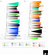
Neighbor-joining tree showing the family-specific amplification of NBS sequences. (a) TNLs. (b) CNLs. The complete tree was based on 1,600 sequences (see Additional data files 1 and 2 for an expanded tree with individual sequences and the alignments used). Clades that contained sequences from individual plant families were collapsed into single branches and the number of sequences in each branch is indicated. Different taxa are assigned different colors; clades with representatives from several families are shown in black. The scale bar represents five nucleotide substitutions.
Figure 3
Figure 3
Predicted structures of NBS domains. Structural models for the NBS domain of TNL RPS4 and CNL RPS5 of Arabidopsis were generated using a self-consistent mean-field homology modeling technique [95], in the absence of ADP. ADP was added to the two NBS models by inference from the APAF-1-ADP complex without further refinement of the models to illustrate the position of the nucleotide relative to the conserved motifs. (a)The structures of the NBS domains of RPS4 and RPS5, showing the positions of the conserved motifs. The protein structures are shown as ribbon diagrams and ADP is shown as a stick model. TIR-type and CC-type NBS domains are made up of motifs, in order from the amino terminus [3]: P-loop (or Walker A site, blue); RNBS-A (green); kinase-2 (or Walker B site, magenta); RNBS-B (green); RNBS-C (green); GLPL (yellow); RNBS-D (green); MHDV (orange). (b) The binding sites of human APAF-1 (PDB code 1z6tA), Arabidopsis RPS4, and RPS5, showing the residues interacting with ADP and ATP. The coordination of ADP in the three proteins involves three different conserved motifs. A small anchor region at the amino terminus of the NBS domain coordinates the adenine of ADP or ATP, the P-loop coordinates the α- and β-phosphates, and the MHDV motif (in the winged-helix subdomain in APAF-1) coordinates either the sugar or the β-phosphate of ADP. The two terminal aspartic acids from the kinase-2 motif are located in the pocket in which the γ-phosphate of ATP would sit. Images were generated using PyMol [96].
Figure 4
Figure 4
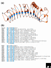
The predicted structure of an LRR domain resulting from the threading of the LRR domain of Arabidopsis RPS5 onto bovine decorin (PDB code 1xku). (a) A cartoon representation of the predicted structure of the RPS5 LRR domain generated using PyMol [96]. The β-sheets forming the concave face of the 'horseshoe' are represented as arrows. The conserved aliphatic residues are shown in blue. N, amino terminus; C, carboxyl terminus. (b) Alignment of the 12 leucine-rich repeats in decorin and the 13 repeats in RPS5 as well as the amino terminal nine amino acids. The conserved aliphatic residues are shown in blue.
Figure 5
Figure 5
The regulatory interactions between two Arabidopsis CNL proteins, RPM1 and RPS2, RPM1-Interacting Protein 4 (RIN4) and three pathogen virulence effectors, AvrB, AvrRpm1 and AvrRpt2 [51-55]. The protein RPM1 detects the phosphorylation of RIN4 by AvrB and AvrRpm1 and elicits the resistance response. This outcome can be blocked by AvrRpt2, a protease that cleaves RIN4. The disappearance of RIN4 is detected by RPS2, resulting in elicitation of the defense response.

Similar articles

Cited by

References

    1. Jones DA, Jones JDG. The role of leucine-rich repeat proteins in plant defenses. Adv Bot Res. 1997;24:90–167.
    1. Ellis J, Dodds P, Pryor T. Structure, function and evolution of plant disease resistance genes. Curr Opin Plant Biol. 2000;3:278–284. doi: 10.1016/S1369-5266(00)00080-7. - DOI - PubMed
    1. Meyers BC, Kozik A, Griego A, Kuang H, Michelmore RW. Genome-wide analysis of NBS-LRR-encoding genes in Arabidopsis. Plant Cell. 2003;15:809–834. doi: 10.1105/tpc.009308. - DOI - PMC - PubMed
    1. Belkhadir Y, Subramaniam R, Dangl JL. Plant disease resistance protein signaling: NBS-LRR proteins and their partners. Curr Opin Plant Biol. 2004;7:391–399. doi: 10.1016/j.pbi.2004.05.009. - DOI - PubMed
    1. Jones DA, Takemoto D. Plant innate immunity - direct and indirect recognition of general and specific pathogen-associated molecules. Curr Opin Immunol. 2004;16:48–62. doi: 10.1016/j.coi.2003.11.016. - DOI - PubMed