

doi: 10.1016/j.canlet.2006.03.029.
Epub 2006 May 4.
Development of a CA125-mesothelin cell adhesion assay as a screening tool for biologics discovery
Affiliations
- PMID: 16677756
- PMCID: PMC2734268
- DOI: 10.1016/j.canlet.2006.03.029
Item in Clipboard
Development of a CA125-mesothelin cell adhesion assay as a screening tool for biologics discovery
Cancer Lett.
.
Abstract
Preventing peritoneal implantation of ovarian carcinoma cells could prolong patient remission and survival. CA125 is expressed on most ovarian cancer cells and was reported to be a ligand of mesothelin, a peritoneal protein. We developed a cell adhesion assay with CA125-expresser ovarian cancer cells and human mesothelin-transfected cells and we confirmed that CA125 and mesothelin mediate cell attachment. We also showed that this assay supplies a high-throughput screening system for reagents able to block CA125/mesothelin-dependent cell attachment with a sensitive quantitative readout. We finally demonstrated that a mesothelin chimeric protein and anti-CA125 antibodies block CA125/mesothelin-dependent cell attachment.
Figures
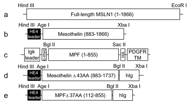
Constructs. (a)–(c) Constructs for cell surface expression: (a) full-length MSLN1 cDNA (1866 bp) was ligated to pcDNA3.1/−zeo(+) vector in between Hind III and EcoR I sites; (b) the N-terminal domain of MSLN1 cDNA that encodes mesothelin (984 bp) was ligated to a modified pcDNA3.1/hygro(+) vector in between Age I and Xba I sites and in frame with an HE4 leader sequence; (c) the C-terminal domain of MSLN1 cDNA that encodes MPF (855 bp) was ligated to pDisplay vector between Bgl II and Sac II sites and in frame with Igk leader sequence, c-myc tag, HA tag and PDGFR transmembrane domain. (d)–(e) Constructs for secreted chimeric proteins: truncated forms of (d) mesothelin (855 bp) and (e) MPF (744 bp) were ligated to a modified pcDNA3.1/hygro(+) vector between Age I and Bgl II sites and in frame with HE4 leader and human Ig sequence. The beginning and end positions of the inserts relative to the sequence of MSLN1 clone IMAGE no. 3957372 are shown in parenthesis. The number of truncated amino acids (AA) follows the Δ symbol.
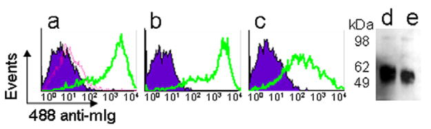
Validation of transfected HEK293F cells. (a)–(c) Flow cytometry analysis: HEK293F cells transfected with (a) MSLN1 or (b) mesothelin constructs were incubated with 10 μg/ml of 4H3 mAb (solid lines); (c) HEK293F cells transfected with MPF construct were incubated with 10 μg/ml of anti-c-myc tag mAb (solid lines). As negative controls, wild type (dotted line) or transfected cells (shade areas) were incubated with the secondary antibody only. (d)–(e) Western blot: (d) Meso-Ig and (e) MPF-Ig chimeric proteins were detected with an HRP-anti-hIg mAb.
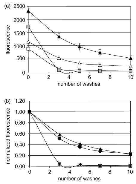
CA125/mesothelin-dependant cell adhesion assay. Ninety six-well plates containing OVCAR-3 adherent cells and fluorescently-labeled HEK 293 cells, wild type at 2.5×10⁶ cells/ml (white squares) or 5×10⁶ cells/ml (gray squares); or transfected with mesothelin at 2.5×10⁶ cells/ml (white triangles) or 5×10⁶ cells/ml (black triangles); MPF at 5×10⁶ cells/ml (black diamonds); MSLN1 at 5×10⁶ cells/ml (black circles) were washed up to 10 times. (a) Remaining fluorescence was plotted after each wash or (b) normalized to the original fluorescence for each well. The assay was performed in triplicates and the values were averaged. All standard deviations were less than 4%.
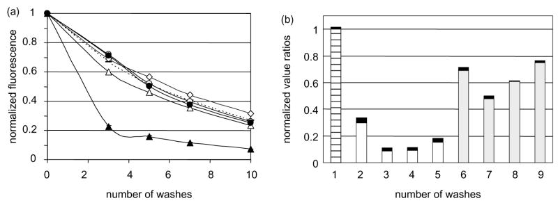
Cross-linked Meso-Ig and anti-CA125 mAb block cell adhesion (a) The cell adhesion assay was conducted in presence of 10 μg/ml of MPF-Ig alone (white circles) or cross-linked with anti-Ig mAb (black circles), or Meso-Ig alone (white triangles) or cross linked (black triangles). As a positive control for cell attachment, the assay was performed in medium only (white diamonds) or in medium supplemented with anti-hIg (dotted line). The assays were performed in triplicates. All standard deviations were less than 4%. (b) The cell adhesion assay was conducted after preincubation of OVCAR3 cells with 10 μg/ml of anti-CA125 antibodies (2=M002203; 3=M11; 4=M8072321; 5=X52; 6=M002201; 7=OC-125; 8=M8072320; 9=X306). The normalized fluorescent signals after five washes were divided by the fluorescent signal of the adhesion assay performed in medium only (1). The gray bars correspond to the anti-CA125 mAb of group A and the white bars to the group B. The black boxes represent the standard deviations and the stripped bar is the positive control for cell attachment.
References
-
- Rump A, Morikawa Y, Tanaka M, Minami S, Umesaki N, Takeuchi M, et al. Binding of ovarian cancer antigen CA125/MUC16 to mesothelin mediates cell adhesion. J Biol Chem. 2004;279(10):9190–9198. - PubMed
-
- Yamaguchi N, Yamamura Y, Konishi E, Ueda K, Kojima T, Hattori K, et al. Characterization, molecular cloning and expression of megakaryocyte potentiating factor. Stem Cells. 1996;14(Suppl 1):62–74. - PubMed
-
- Kojima T, Oheda M, Hattori K, Taniguchi Y, Tamura M, Ochi N, et al. Molecular cloning and expression of megakaryocyte potentiating factor cDNA. J Biol Chem. 1995;270(37):21984–21990. - PubMed
Publication types
MeSH terms
Substances
Associated data
- Actions
- Actions
Grants and funding
LinkOut - more resources
Full Text Sources
Other Literature Sources
Medical
Research Materials
Miscellaneous
