. 2006 Aug 1;351(2):291-302.
doi: 10.1016/j.virol.2006.03.042. Epub 2006 May 6.

Core labeling of adenovirus with EGFP

Affiliations

Core labeling of adenovirus with EGFP

Long P Le et al. Virology. .

Abstract

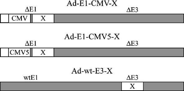
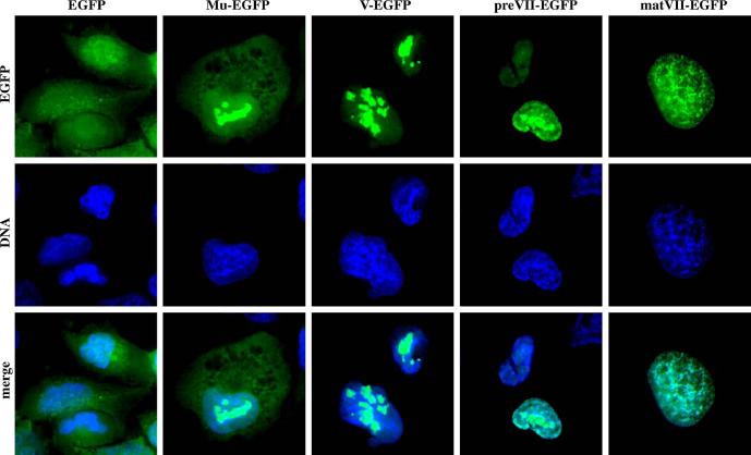
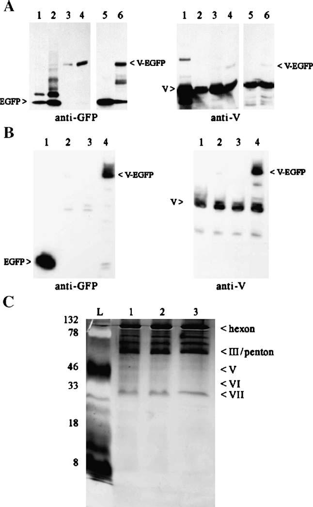
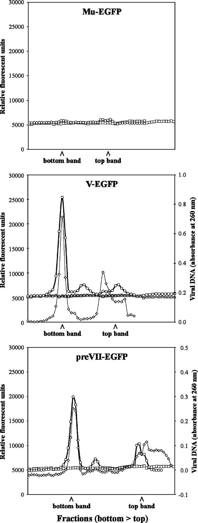
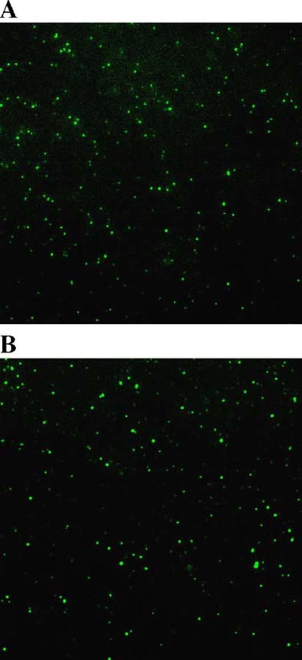
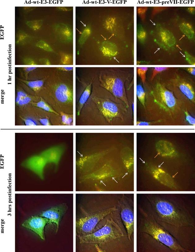
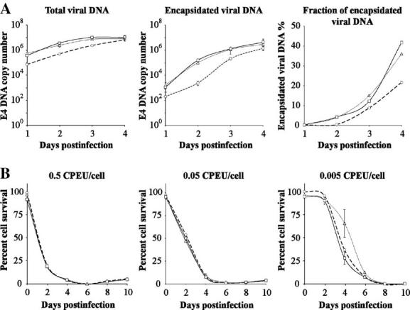
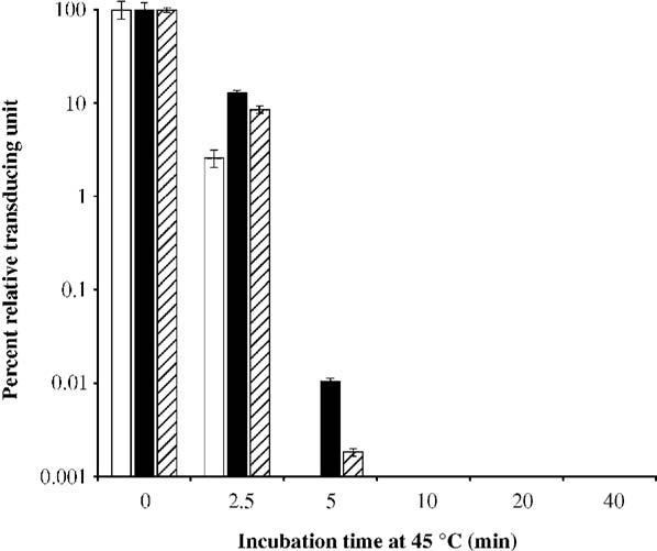
The study of adenovirus could greatly benefit from diverse methods of virus detection. Recently, it has been demonstrated that carboxy-terminal EGFP fusions of adenovirus core proteins Mu, V, and VII properly localize to the nucleus and display novel function in the cell. Based on these observations, we hypothesized that the core proteins may serve as targets for labeling the adenovirus core with fluorescent proteins. To this end, we constructed various chimeric expression vectors with fusion core genes (Mu-EGFP, V-EGFP, preVII-EGFP, and matVII-EGFP) while maintaining expression of the native proteins. Expression of the fusion core proteins was suboptimal using E1 expression vectors with both conventional CMV and modified (with adenovirus tripartite leader sequence) CMV5 promoters, resulting in non-labeled viral particles. However, robust expression equivalent to the native protein was observed when the fusion genes were placed in the deleted E3 region. The efficient Ad-wt-E3-V-EGFP and Ad-wt-E3-preVII-EGFP expression vectors were labeled allowing visualization of purified virus and tracking of the viral core during early infection. The vectors maintained their viral function, including viral DNA replication, viral DNA encapsidation, cytopathic effect, and thermostability. Core labeling offers a means to track the adenovirus core in vector targeting studies as well as basic adenovirus virology.


Figures

Fig. 1
Fig. 1
Schematic of vectors. Nomenclature and configuration of expression vectors where X represents Mu-EGFP, V-EGFP, preVII-EGFP, or matVII-EGFP. The employed expression strategies include transcription via the typical CMV promoter, a modified CMV promoter containing the adenovirus tripartite leader sequence (CMV5), and the adenovirus major late promoter (by virtue of transgene cassette insertion into the deleted E3 region).
Fig. 2
Fig. 2
Fluorescent core fusion protein expression and localization. A549 cells were infected with nonreplicative E1-CMV adenovirus vectors expressing the indicated genes. The cells were fixed and stained for nuclear DNA 24 h postinfection followed by visualization of the fluorescent proteins by epifluorescence microscopy.
Fig. 3
Fig. 3
Western blot analysis of Ad-E1-CMV-V-EGFP purified virus and cells infected with various expression vectors. (A) Purified viruses (8 μg protein in each sample) and infected cell lysates (4 μg protein each sample) were subjected to SDS-PAGE and blotted with GFP and pV antibodies. Lanes (1) Ad-E1-CMV-EGFP bottom band, (2) Ad-E1-CMV-EGFP top band, (3) Ad-E1-CMV-V-EGFP bottom band, (4) Ad-E1-CMV-V-EGFP top band, (5) Ad-E1-CMV-GFP infected 911 cells, and (6) Ad-E1-CMV-V-EGFP infected 911 cells. (B) Protein lysates (10 μg) from 911 cells infected with the various V-EGFP expression vectors were subjected to SDS-PAGE and blotted with GFP and pV antibodies. Lanes (1) Ad-E1-CMV-EGFP, (2) Ad-E1-CMV-V-EGFP, (3) Ad-E1-CMV5-V-EGFP, and (4) Ad-wt-E3-V-EGFP. (C) Purified core-labeled viral particles (1.5 × 10¹⁰) were subjected to SDS-PAGE and then silver-stained. Lanes: (L) protein ladder, (1) Ad-wt-E3-EGFP, (2) Ad-wt-E3-V-EGFP, and (3) Ad-wt-E3-preVII-EGFP. The numbers on the left side indicate the molecular weight of the protein ladder (kDa). The labels on the right highlight the major Ad structural proteins visible on the SDS-PAGE.
Fig. 4
Fig. 4
Analysis of fusion core protein viral gradients with improved expression. Fusion core protein vectors with enhanced expression were purified and fractionated to determine fluorescence of CsCl gradient fractions. For all panels, unlabeled Ad-E1-CMV5-EGFP was included as a control (–□–). Ad-E1-CMV5-X (–sh=utri–) and Ad-wt-E3-X (–○–) are shown where X is the indicated fusion protein at the top of the panel. Note that Ad-E1-CMV5-Mu-EGFP, Ad-E1-CMV5-preVII-EGFP, Ad-E1-CMV5-matVII-EGFP, and Ad-wt-E3-matVII-EGFP could not be rescued for analysis. For Ad-wt-E3-V-EGFP and Ad-wt-E3-preVII-EGFP, viral DNA content was also analyzed for the respective fractions (–◇–).
Fig. 5
Fig. 5
Visualization of Ad-wt-E3-V-EGFP and Ad-wt-E3-preVII-EGFP particles. Purified Ad-wt-E3-V-EGFP (A) and Ad-wt-E3-preVII-EGFP (B) were prepared on slides with coverslips and imaged with epifluorescence microscopy.
Fig. 6
Fig. 6
Tracking of core-labeled virus infection. HeLa cells were infected with Ad-wt-E3-EGFP (left column), Ad-wt-E3-V-EGFP (middle column), and Ad-wt-E3-preVII-EGFP (right column) at 37 °C and then imaged at 1 h (upper 2 rows) and 3 h (bottom 2 rows) postinfection using epifluorescence microscopy. Green represents EGFP fluorescence and some background autofluorescence; blue indicates nuclear DNA; and red, yellow, and brown represent background signal. White arrows = fluorescent viral particles in the cytoplasm. Orange arrows = fluorescent viral particles in the nucleus. Note that the bottom row pictures for each time period are merged color images overlaid on top of a phase contrast picture of the cells to show morphology.
Fig. 7
Fig. 7
DNA packaging efficiency and cytopathic effect of core-labeled viruses. (A) 911 cells were infected over the course of 4 days with control Ad-wt-E3-EGFP (--○--), Ad-wt-E3-V-EGFP (–△–), and Ad-wt-E3-preVII-EGFP (–□–). Each day, half of the cells collected were processed for total viral DNA while the other half were processed for encapsidated viral DNA. Both viral DNA pools were quantitated by Taqman real-time quantitative PCR using E4-specific primers (n = 3). The third panel shows the percent of encapsidated viral DNA for each respective virus (encapsidated divided by total and then multiplied by 100%). (B) The cytopathic effect of the same three viruses was analyzed in 911 cells over the course of 10 days using the indicated multiplicities of infection. Every 2 days, the cell viability was quantitated by MTS assay. Cell viability is expressed as percentage relative to noninfected cells (n = 5). Ad-wt-E3-EGFP (--○--), Ad-wt-E3-V-EGFP (–△–), and Ad-wt-E3-preVII-EGFP (–□–).
Fig. 8
Fig. 8
Thermostability of core-labeled viruses. Ad-wt-E3-EGFP (white), Ad-wt-V-EGFP (black), and Ad-wt-E3-preVII-EGFP (striped) were incubated at 45 °C for various times and then quantitated by transduction unit titer. Bars represent percent of remaining infectivity relative to untreated samples (n = 3).

References

    1. Alemany R, Suzuki K, Curiel DT. Blood clearance rates of adenovirus type 5 in mice. J. Gen. Virol. 2000;81(Pt 11):2605–2609. - PubMed
    1. Alestrom P, Akusjarvi G, Lager M, Yeh-kai L, Pettersson U. Genes encoding the core proteins of adenovirus type 2. J. Biol. Chem. 1984;259(22):13980–13985. - PubMed
    1. Anderson CW, Young ME, Flint SJ. Characterization of the adenovirus 2 virion protein, mu. Virology. 1989;172(2):506–512. - PubMed
    1. Berk AJ. Adenovirus promoters and E1A transactivation. Annu. Rev. Genet. 1986;20:45–79. - PubMed
    1. Berkner KL, Sharp PA. Effect of the tripartite leader on synthesis of a non-viral protein in an adenovirus 5 recombinant. Nucleic Acids Res. 1985;13(3):841–857. - PMC - PubMed

Publication types

Substances