

Image analysis and superimposition of 3-dimensional cone-beam computed tomography models
- PMID: 16679201
- PMCID: PMC3586191
- DOI: 10.1016/j.ajodo.2005.12.008
Image analysis and superimposition of 3-dimensional cone-beam computed tomography models
Abstract
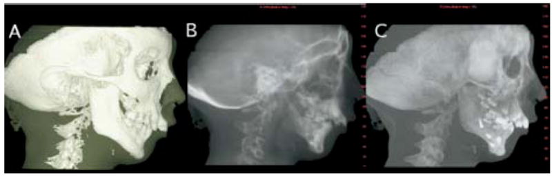
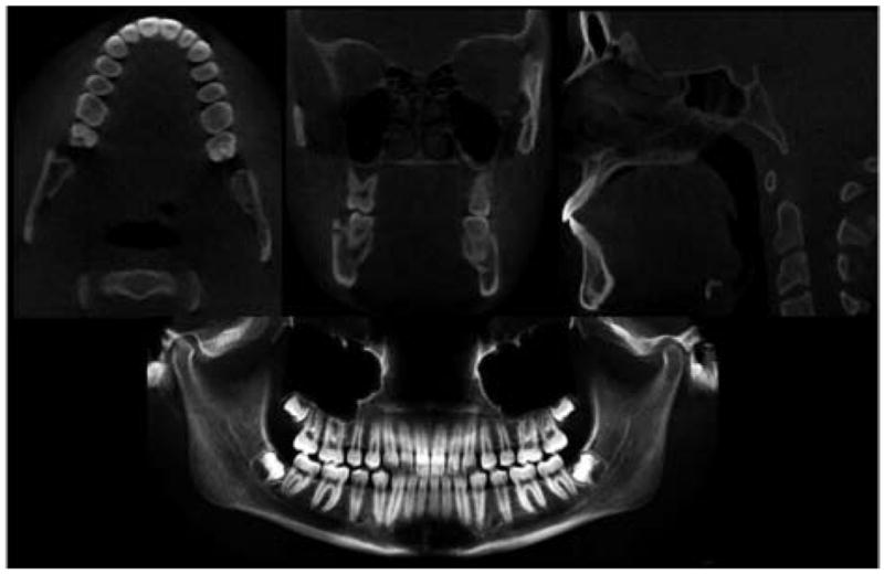
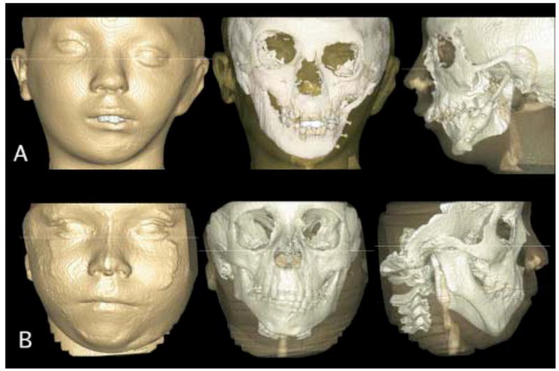
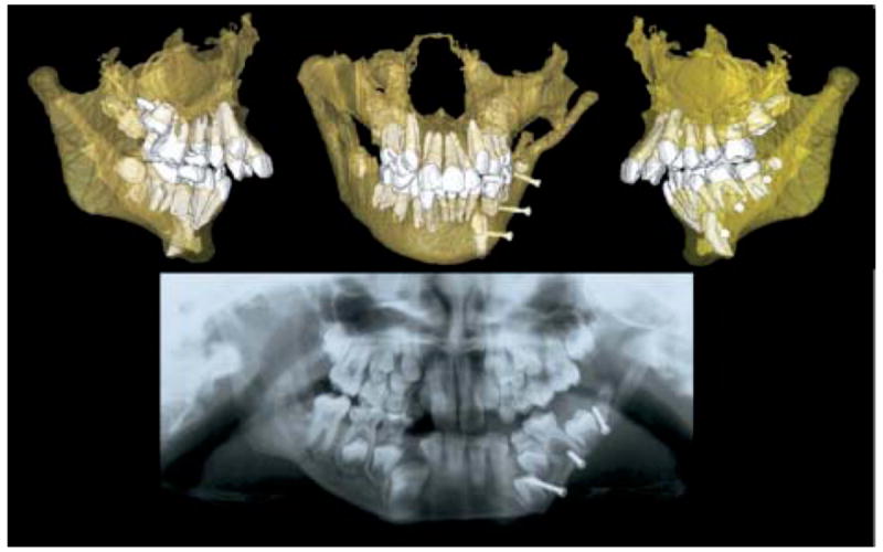
Three-dimensional (3D) imaging techniques can provide valuable information to clinicians and researchers. But as we move from traditional 2-dimensional (2D) cephalometric analysis to new 3D techniques, it is often necessary to compare 2D with 3D data. Cone-beam computed tomography (CBCT) provides simulation tools that can help bridge the gap between image types. CBCT acquisitions can be made to simulate panoramic, lateral, and posteroanterior cephalometric radioagraphs so that they can be compared with preexisting cephalometric databases. Applications of 3D imaging in orthodontics include initial diagnosis and superimpositions for assessing growth, treatment changes, and stability. Three-dimensional CBCT images show dental root inclination and torque, impacted and supernumerary tooth positions, thickness and morphology of bone at sites of mini-implants for anchorage, and osteotomy sites in surgical planning. Findings such as resorption, hyperplasic growth, displacement, shape anomalies of mandibular condyles, and morphological differences between the right and left sides emphasize the diagnostic value of computed tomography acquisitions. Furthermore, relationships of soft tissues and the airway can be assessed in 3 dimensions.
Figures
References
-
- Halazonetis DJ. From 2-dimensional cephalograms to 3-dimensional computed tomography scans. Am J Orthod Dentofacial Orthop. 2005;127:627–37. - PubMed
-
- Hatcher DC, Aboudara CL. Diagnosis goes digital. Am J Orthod Dentofacial Orthop. 2004;125:512–5. - PubMed
-
- Mah J, Hatcher DC. Three-dimensional craniofacial imaging. Am J Orthod Dentofacial Orthop. 2004;126:308–9. - PubMed
-
- Hajeer MJ, Ayoub AF, Millett DT, Bock M, Siebert JP. Three-dimensional imaging in orthognathic surgery: the clinical application of a new method. Int J Adult Orthod Orthognath Surg. 2002;17:318–30. - PubMed
-
- Chirani RA, Jacq JJ, Meriot P, Roux C. Temporomandibular joint: a methodology of magnetic resonance imaging 3-D reconstruction. Oral Surg Oral Med Oral Pathol Radiol Endod. 2004;97:756–61. - PubMed
Publication types
MeSH terms
Grants and funding
LinkOut - more resources
Full Text Sources
Other Literature Sources
Medical
Miscellaneous
