. 2006 Jun;18(6):1467-76.
doi: 10.1105/tpc.106.042036. Epub 2006 May 5.

Overexpression of INFLORESCENCE DEFICIENT IN ABSCISSION activates cell separation in vestigial abscission zones in Arabidopsis

Affiliations

Overexpression of INFLORESCENCE DEFICIENT IN ABSCISSION activates cell separation in vestigial abscission zones in Arabidopsis

Grethe-Elisabeth Stenvik et al. Plant Cell. 2006 Jun.

Abstract

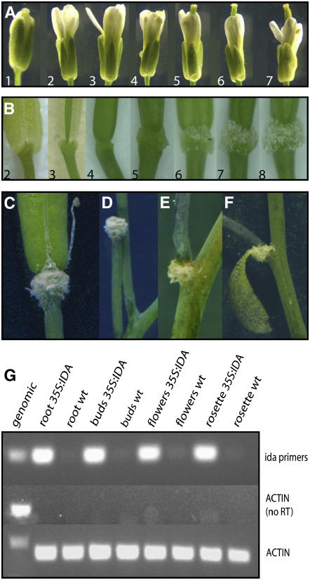
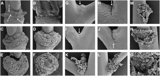
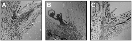
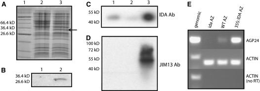
Plants may shed organs when they have been injured or served their purpose. The differential pattern of organ abscission in different species is most likely the result of evolutionary adaptation to a variety of life styles and environments. The final step of abscission-related cell separation in floral organs of wild-type Arabidopsis thaliana, which only abscises sepals, petals, and stamens, is controlled by INFLORESCENCE DEFICIENT IN ABSCISSION (IDA). Here, we demonstrate that Arabidopsis 35S:IDA lines constitutively overexpressing IDA exhibit earlier abscission of floral organs, showing that the abscission zones are responsive to IDA soon after the opening of the flowers. In addition, ectopic abscission was observed at the bases of the pedicel, branches of the inflorescence, and cauline leaves. The silique valves also dehisced prematurely. Scanning electron microscopy indicated a spread of middle lamella degradation from preformed abscission zone cells to neighboring cells. A transcript encoding an arabinogalactan protein (AGP) was upregulated in the 35S:IDA lines, and large amounts of AGP were secreted at the sites of abscission. AGP was shown to be a constituent of wild-type floral abscission zones during and soon after cell separation had been completed. We suggest that the restricted expression pattern of IDA precludes abscission of nonfloral organs in Arabidopsis.


Figures

Figure 1.
Figure 1.
Phenotype of 35S:IDA Plants. (A) Flowers and siliques of a wild-type plant from positions 1 to 7 along the inflorescence, as indicated. (B) Flowers and siliques of a 35S:IDA plant from positions 2 to 8 along the inflorescence, as indicated. Position 3, abscised sepals; position 4, abscised sepals, petals, and stamens; position 5, enlarged AZ region; positions 6 to 8, secreted white substance. (C) Full-grown green silique with the white substance covering the AZ, premature opening of the valves, and secretion of the white substance along the dehiscence zone. (D) Pedicel after shedding of the silique. (E) Secretion of the white substance and abscission at the base of a branch. (F) Secretion of the white substance at the base of a cauline leaf about to abscise. (G) RT-PCR analysis of different tissues from 35S:IDA and wild-type plants, as indicated, using primers amplifying a 237-bp IDA fragment. Primers for ACTIN2-7 were used to amplify a 255-bp fragment from all tissues as a positive control or a 340-bp fragment from genomic DNA. Actin without RT was used as a negative control.
Figure 2.
Figure 2.
Scanning Electron Micrographs of Floral AZs and the Base of the Pedicel. (A) Wild-type AZ of sepals (S), petals (P), and filaments (F) after abscission, as indicated. (B) Mutant ida AZ with broken cells after forcible removal of floral organs. (C) Enlarged 35S:IDA AZs. (D) 35S:IDA AZ with a dramatically greater number of rounded cells. (E) Gynophore detached from the 35S:IDA AZ. (F) 35S:IDA AZ showing broken cells where the gynophore was attached. (G) Wild-type pedicel vestigial AZ with a band of small cells (arrow). (H) Wild-type pedicel vestigial AZ. A small cleft (arrow) developed in older pedicels. (I) and (J) Pedicel AZs in 35S:IDA plants at stages comparable to the wild type shown in (G) and (H). (K) and (L) Pedicel AZs in 35S:IDA plants, later stages. (M) Base of the pedicel in a 35S:IDA plant showing rounded cells after abscission of the pedicel. (N) AZ showing rounded cells after shedding of the cauline leaf in a 35S:IDA plant. (O) Close-up of the cells in the cauline leaf AZ.
Figure 3.
Figure 3.
Light Microscopic Analysis of Thin Sections of the Regions at the Bases of Organs in Wild-Type AZ Cells (Arrows). (A) Branching point. (B) Base of a cauline leaf. (C) Base of a pedicel.
Figure 4.
Figure 4.
Identification of Arabinogalactan by the Yariv Reagent β-GlcY. (A) to (F) Staining of siliques by β-GlcY, resulting in a red precipitate. Positions are numbered. The bottom row shows greater magnifications of the stained AZs. (G) Mature silique stained with β-GlcY. (H) Mature silique stained with the negative control α-GlcY. (I) Position-10 silique of a wild-type plant stained with β-GlcY. (J) Separating region of a 35S:IDA filament stained with β-GlcY.
Figure 5.
Figure 5.
IDA and AGP Expression in AZs. (A) Coomassie blue–stained SDS-PAGE gel of proteins from E. coli cultures harboring a GST:IDA fusion construct, with and without induction with isopropylthio-β-galactoside (lanes 2 and 3, respectively). The induced fusion protein is indicated by an arrow. The sizes of relevant markers of the protein standard (lane 1) are shown at left. (B) Detection of the GST-IDA fusion protein by protein gel blot analysis of proteins from uninduced (lane 1) or induced (lane 2) E. coli cells using the IDA antibody. (C) Protein gel blot analysis of 200 μg of protein isolated from AZ tissue of wild-type (lane 1), ida mutant (lane 2), and 35S:IDA (lane 3) plants using the IDA antibody (Ab). Size markers are shown at left. (D) The same membrane as in (C) probed with JIM13. (E) RT-PCR analysis with AGP24 primers of cDNA derived from AZs of ida, wild-type (ecotype C24), and 35S:IDA plants, as indicated. Actin primers were used for positive and negative controls with both genomic DNA and cDNA.

References

    1. Aalen, R.B., Butenko, M.A., Stenvik, G.-E., Tandstad, N.M., and Patterson, S.E. (2006). Genetic control of floral abscission. In Floriculture, Ornamental and Plant Biotechnology: Advances and Topical Issues, Vol. 1, J.T. da Silva, ed (London: Global Science Books), pp. 101–108.
    1. Addicott, F.T. (1982). Abscission. (London: University of California Press).
    1. Alsop, D.W. (1974). Rapid single-solution polychrome staining of semithin epoxy sections using polyethylene glycol 200 (PEG 200) as a stain solvent. Stain Technol. 49 265–272. - PubMed
    1. Bleecker, A., and Patterson, S.E. (1997). Last exit: Senescence, abscission, and meristem arrest in Arabidopsis. Plant Cell 9 1169–1179. - PMC - PubMed
    1. Borner, G.H., Lilley, K.S., Stevens, T.J., and Dupree, P. (2003). Identification of glycosylphosphatidylinositol-anchored proteins in Arabidopsis. A proteomic and genomic analysis. Plant Physiol. 132 568–577. - PMC - PubMed

Publication types

LinkOut - more resources