. 2006 Apr;47(4):578-86.
doi: 10.1097/01.fjc.0000211740.47960.8b.

Constitutively active Src tyrosine kinase changes gating of HCN4 channels through direct binding to the channel proteins

Affiliations

Constitutively active Src tyrosine kinase changes gating of HCN4 channels through direct binding to the channel proteins

Suzanne S Arinsburg et al. J Cardiovasc Pharmacol. 2006 Apr.

Abstract

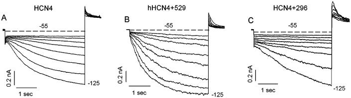
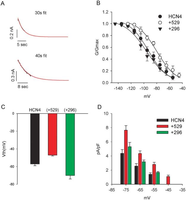
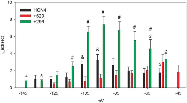
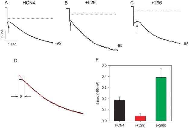
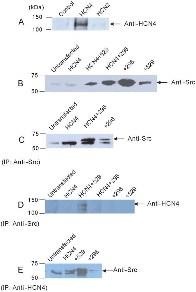
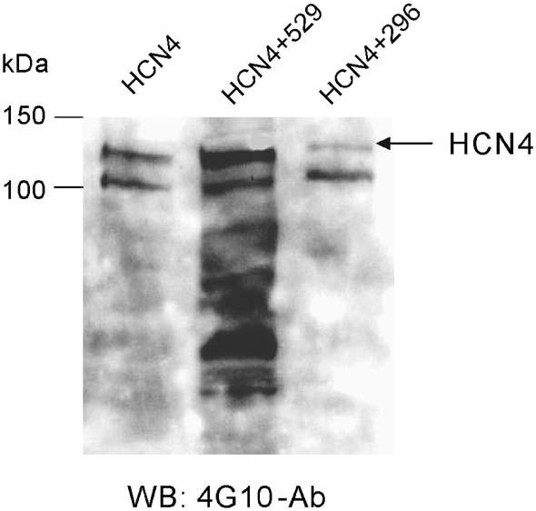
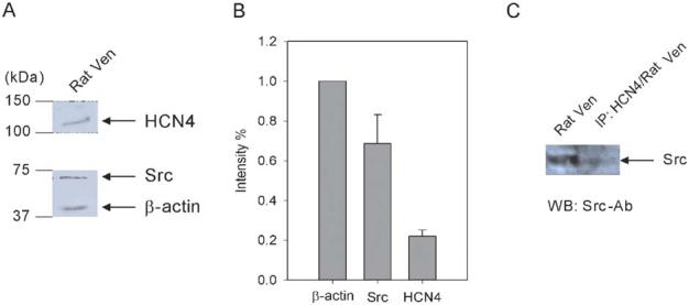
Cardiac pacemaker current, if, is generated by hyperpolarization-activated cyclic nucleotide-gated (HCN) channels. Our previous studies demonstrated that altered tyrosine phosphorylation can modulate the properties of both if and HCN channels. To assess a hypothesis that the intracellular tyrosine kinase Src may play a role in modulation by tyrosine phosphorylation of if, we cotransfected HEK293 cells with HCN4 and Src proteins. When HCN4 was cotransfected with a constitutively activated Src protein (Src529), the resultant voltage-dependent HCN4 activation was positively shifted (HCN4: V1/2 = -93 mV; Src529: V1/2 = -80 mV). The activation kinetics were accelerated at some potentials but not over the entire voltage range tested (eg, at -95 mV, tau_act(HCN4) = 3,243 ms; tau_act(Src529) = 1,113 ms). When HCN4 was cotransfected with a dominant negative Src protein (Src296), the HCN4 activation was shifted more negative to a smaller degree (HCN4: V1/2 = -93 mV; Src296: V1/2 = -98 mV; statistically insignificant) and the activation kinetics were slowed at most test potentials (eg, at -95 mV, tau_act(Src296) = 7,396 ms). Neither Src529 nor Src296 significantly altered HCN4 current density. Coimmunoprecipitation experiments revealed that Src forms a complex with HCN4 in HEK293 cells and in rat ventricular myocytes. Our data provide a novel mechanism of if regulation by Src tyrosine phosphorylation.


Figures

FIGURE 1
FIGURE 1
An example of expression of HCN4 in HEK293 cells. From the holding potential of −10 mV, the cells were typically hyperpolarized to potentials ranging from −55 to −125 mV in 10 mV increment. Currents shown are for HCN4 alone (A), HCN4+Src529 (B), and HCN4+Src296 (C). The dashed lines represent the 0 currents.
FIGURE 2
FIGURE 2
HCN4 activation. A, Method of fitting to obtain steady-state estimates of current activation (see text for details). B, Activation curves for HCN4, HCN4+Src529, and HCN4+Src296. The mean midpoints and slope factors of activation for HCN4 (−93 mV, −16 mV) and HCN4+Src529 (−80 mV, −16 mV) and HCN4+Src296 (−98 mV, −18 mV) were used to generate the fitting curves. C, Mean voltage thresholds for activation of HCN4, HCN4+Src529, and HCN4+Src296. D, Current density expressed as pA/pF at potentials in the range of −45 to −75 mV. There is more current with Src529 than HCN4 or HCN4+Src296 in this voltage range.
FIGURE 3
FIGURE 3
Activation kinetics of HCN4, HCN4+Src529, and HCN4+Src296. Activation kinetics were obtained by fitting the onset current traces to 30 or 40 s. Averaged kinetics data are presented from 8 cells expressing HCN4 and HCN4+Src529 and 10 cells expressing HCN4+296. Numbers above the bars indicate the number of cells analyzed at those potentials. #P <0.05 for HCN4 and HCN4+Src296 comparison; &P <0.05 for HCN4 and HCN4+Src529 comparison.
FIGURE 4
FIGURE 4
Initial delays (arrows) in HCN4 current activation at −95 mV. A typical set of current traces at −95 mV are shown for HCN4 (A), HCN4+Src529 (B), and HCN4+Src296 (C). Dotted lines are zero currents. D, Measurement of the initial delay: Delay is defined as the time difference between the beginning of current response to the voltage pulse and the beginning of the current trace that can be best fit by a single exponential function; labeled with the symbol δ. The capacitance current was excluded from the measurement. E, Mean delays are 0.185 ± 0.033(s) for HCN4 (n = 8), 0.044 ± 0.021(s) for HCN4+Src529 (n = 8), and 0.391 ± 0.078(s) for HCN4+Src296 (n = 10).
FIGURE 5
FIGURE 5
Immunoprecipitation of HCN4 with Src proteins in HEK cells. A, Western blot of HCN4 in untransfected, HCN4, and HCN2 cells. B, Western blot of Src in untransfected, HCN4, HCN4+Src529, HCN4+Src296, Src296, and Src529 cells. C, IP with anti-Src followed by Western blotting using anti-Src in untransfected, HCN4, HCN4+Src296, and Src296 alone cells. D, IP with anti-Src followed by Western blotting using anti-HCN4 in untransfected, HCN4, HCN4+Src529, HCN4+Src296, Src296, and Src529 cells. E, IP with anti-HCN4 followed by Western blotting using anti-Src in untransfected, HCN4, HCN4+Src529, and HCN4+Src296.
FIGURE 6
FIGURE 6
Tyrosine phosphorylation of HCN4 channels. Phosphorylated tyrosine signals in HEK cells transfected with HCN4, HCN4+Src529, and HCN4+Src296 were detected in Western blotting using a phosphotyrosine specific antibody, 4G10.
FIGURE 7
FIGURE 7
Interaction of HCN4 with Src in rat ventricle. A, Western blots of HCN4 and Src proteins in rat ventricle. The membranes were cut in half. The two halves were incubated with anti-HCN4 and anti-Src antibodies, respectively. The half membrane fraction incubated with anti-Src antibody was washed and reprobed with anti-β-actin antibody to reveal the β-actin signal. B, Src and HCN4 expression levels are normalized to β-actin, which is used as an internal control (n = 3). C, Right lane: rat ventricular sample was immunoprecipitated by a specific HCN4 antibody followed by Western blotting using a specific Src antibody; left lane: Western blotting of rat ventricle sample using a specific Src antibody.

References

    1. Davis MJ, Wu X, Nurkiewicz TR, et al. Regulation of ion channels by protein tyrosine phosphorylation. Am J Physiol Heart Circ Physiol. 2001;281:H1835–H1862. - PubMed
    1. Wu JY, Cohen IS. Tyrosine kinase inhibition reduces if in rabbit SA myocytes. Pflügers Arch. 1997;434:509–514. - PubMed
    1. Wu JY, Yu H, Cohen IS. Epidermal growth factor increases if in rabbit SA node cells by activating a tyrosine kinase. Biochim Biophys Acta. 2000;1463:15–19. - PubMed
    1. Nair BG, Rashed HM, Patel TB. Epidermal growth factor produces inotropic and chronotropic effects in rat hearts by increasing cyclic AMP accumulation. Growth Factors. 1993;8:41–48. - PubMed
    1. Rabkin SW, Sunga P, Myrdal S. The effect of epidermal growth factor on chronotropic response in cardiac cells in culture. Biochem Biophys Res Commun. 1987;146:889–897. - PubMed

MeSH terms