. 2006 Jun;13(6):482-90.
doi: 10.1038/nsmb1093. Epub 2006 May 7.

Exon ligation is proofread by the DExD/H-box ATPase Prp22p

Affiliations

Exon ligation is proofread by the DExD/H-box ATPase Prp22p

Rabiah M Mayas et al. Nat Struct Mol Biol. 2006 Jun.

Abstract

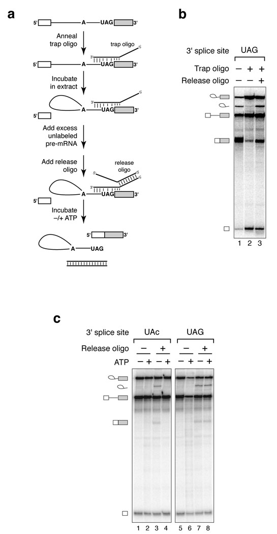
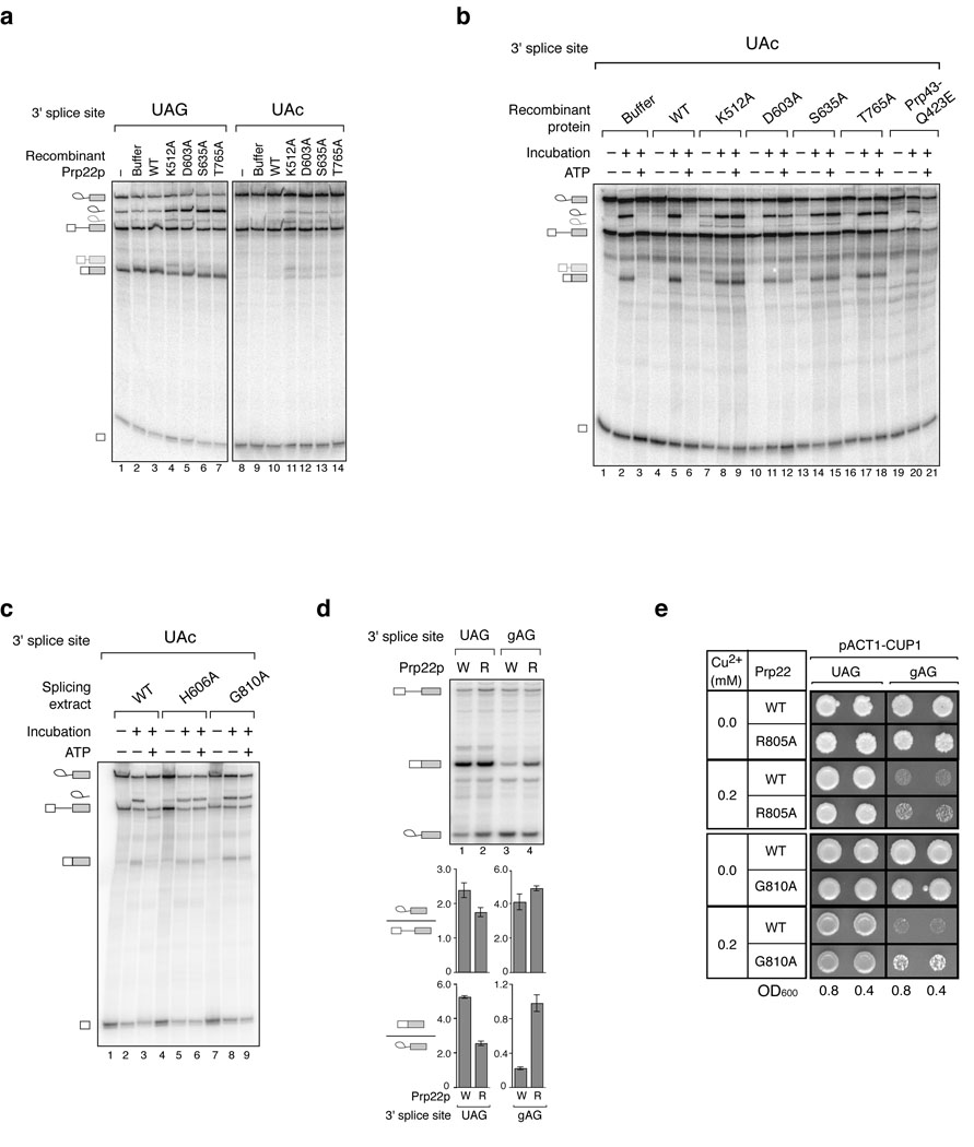
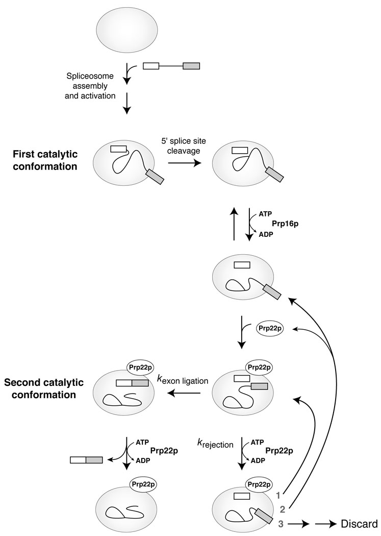
To produce messenger RNA, the spliceosome excises introns from precursor (pre)-mRNA and splices the flanking exons. To establish fidelity, the spliceosome discriminates against aberrant introns, but current understanding of such fidelity mechanisms is limited. Here we show that an ATP-dependent activity represses formation of mRNA from aberrant intermediates having mutations in any of the intronic consensus sequences. This proofreading activity is disabled by mutations that impair the ATPase or RNA unwindase activity of Prp22p, a conserved spliceosomal DExD/H-box ATPase. Further, cold-sensitive prp22 mutants permit aberrant mRNA formation from a mutated 3' splice-site intermediate in vivo. We conclude that Prp22p generally represses splicing of aberrant intermediates, in addition to its known ATP-dependent role in promoting release of genuine mRNA. This dual function for Prp22p validates a general model in which fidelity can be enhanced by a DExD/H-box ATPase.


Figures

Figure 1
Figure 1
An ATP-dependent mechanism represses exon ligation at near-consensus 3’ splice sites. (a) Spliceosomes assembled on pre-mRNA substrates with mutations of the 3’ splice site AG stall at the exon ligation stage (c.f. ref. 23). Spliceosomes were assembled in vitro in budding yeast extract on radiolabeled ACT1 pre-mRNA substrates. Mutations of the 3’ splice site UAG are indicated in lowercase. Migration of the splicing species are indicated to the left and are identical for the wild-type and mutated substrates; from the top: lariat intermediate, excised intron, pre-mRNA, mRNA and cleaved 5’ exon. RNA was separated on a denaturing 6% polyacrylamide gel and visualized by phosphorimager (Molecular Dynamics). (b,c) ATP is required to repress splicing at 3’ splice sites having any single mutation (b), but not double mutations (c), of the conserved 3’ splice site AG. Spliceosomes stalled on substrates having mutations at the 3’ splice site were affinity purified at 4 °C and then either frozen or incubated further at 20 °C with or without nucleotide. (d) Aberrant splicing is repressed by four different NTPs. Spliceosomes stalled on the ACT1 substrate having a UAc 3’ splice site were purified and then frozen (No inc) or incubated further as in b,c.
Figure 2
Figure 2
An ATP-dependent mechanism represses exon ligation at a mutated, but not a wild-type, 3’ splice site. (a) A general strategy to investigate the requirements for 3’ splice site recognition (c.f. refs. –36). (b) Spliceosomes can be trapped reversibly at the stage of 3’ splice site recognition. Wild-type pre-mRNA was annealed with a cognate trap oligonucleotide, incubated in splicing reactions and then further incubated with or without a cognate release oligonucleotide. Untrapped, wild-type pre-mRNA was incubated in parallel. (c) ATP represses splicing at an aberrant 3’ splice site specifically. Mutated (UAc) or wild-type (UAG) pre-mRNA was annealed with a cognate trap oligonucleotide and incubated in splicing reactions. Stalled spliceosomes were affinity purified and then incubated with or without a cognate release oligonucleotide, in the absence or presence of ATP. The oligonucleotides functioned specifically; the UAG and UAc trap and release oligonucleotides were not interchangeable (data not shown).
Figure 3
Figure 3
Repression of aberrant exon ligation at mutated 3’ splice sites is compromised in vitro and in vivo by ATPase- and RNA unwindase-deficient variants of the DEAH-box ATPase Prp22p. (a,b) Recombinant Prp22p variants,– abolish repression of an aberrant 3’ splice site in extract (a) or in affinity-purified spliceosomes (b). Wild-type (UAG) or mutated (UAc) pre-mRNA was incubated in wild-type extract supplemented with nothing (−), buffer, wild-type Prp22p (WT) or mutated Prp22p (amino acid mutations are indicated) in standard splicing reactions. In a, these reactions were analyzed directly; in b, the spliceosomes stalled in these reactions were affinity purified and then treated as in Figure 1b. In b, a Prp43p variant was analyzed in parallel. Note that Prp22p variants block dissociation and subsequent degradation of the excised intron (e.g., ref. 42). Migration of cryptic 3’ splice site cleavage products (see text) are indicated to the left as light gray excised intron and mRNA symbols. (c) Endogenous, Prp22p variants also abolish repression of an aberrant 3’ splice site in purified spliceosomes. Mutated (UAc) pre-mRNA was incubated in reactions with extract of a wild-type PRP22 (WT) strain and the cold-sensitive mutant strains prp22-H606A and prp22-G810A (ref. 42). Spliceosomes stalled in these reactions were immunoprecipitated and then treated as in Figure 1b. The faster migration of excised lariat intron in this gel is due to the concentration of polyacrylamide. (d) A prp22 mutant is defective in discriminating between a wild-type and a mutated 3’ splice site in vivo. A wild-type PRP22 (W) or the mutant prp22-R805A (R) strain was transformed with an ACT1-CUP1 splicing reporter having a wild-type UAG or mutated gAG 3’ splice site. Splicing was analyzed by primer extension. The ratio of mRNA to lariat intermediate is plotted as an indicator of the apparent efficiency of exon ligation; the ratio of lariat intermediate to pre-mRNA is plotted for comparison. The averages and range of values for 2 independent experiments are shown. (e) prp22 mutants are defective in repressing gene expression from an ACT1-CUP1 splicing reporter having the mutated gAG 3’ splice site. A wild-type PRP22 (WT) strain and the mutant strains prp22-R805A (R805A) and prp22-G810A (G810A) were transformed with an ACT1-CUP1 reporter having a wild-type (UAG) or mutated (gAG) 3’ splice site. Cells diluted to 0.8 or 0.4 OD600 were spotted onto plates containing 0.0 or 0.2 mM CuSO4 and grown for 3 days at 33 °C (R805A) or 30 °C (G810A).
Figure 4
Figure 4
An ATP- and Prp22p-dependent mechanism also represses aberrant exon ligation of intermediates having mutations of the branch site or 5’ splice site. (a) Spliceosomes assembled on ACT1 pre-mRNA substrates with the mutated branch site UACUAgC (brG) or the mutated 5’ splice site aUAUGU (G1A) catalyzed formation of the intermediates, albeit inefficiently; formation of mRNA was undetectable. Reactions were performed as in Figure 1a. The wild-type (WT) substrate was spliced in parallel for comparison. The 5’ exon of the G1A substrate is larger than for the other substrates due to differences in the transcription template (see Methods). (b,c) ATP is required to repress exon ligation of aberrant intermediates having the brG (b) or G1A (c) mutations and recombinant Prp22p variants compromise this repression. Reactions were performed as in Figure 3b. Note that affinity purification isolated spliceosomes enriched for lariat intermediate and 5’ exon, indicating that a substantial fraction of pre-mRNA was not engaged in active spliceosomes. In b we confirmed by primer extension that the mutated branch was used as the nucleophile in 5’ splice site cleavage; further, we confirmed by RT-PCR and sequencing of the mRNA that the genuine 5’ splice site and 3’ splice site were utilized (data not shown). In vivo, the G1A mutated 5’ splice site of an ACT1-derived reporter is used exclusively as the electrophile in 5’ splice site cleavage. The asterisk in c indicates the expected migration of mRNA; the low level of G1A lariat intermediate precluded visualization of the mRNA.
Figure 5
Figure 5
A dual role for Prp22p in fidelity and mRNA release suggests a proofreading mechanism– for exon ligation. A simplified splicing pathway is shown. Spliceosome assembly and activation are followed by 5’ splice site cleavage, rearrangement of the spliceosome, exon ligation and mRNA release. The rearrangement is shown in two biochemically separable steps and proceeding through a putative transitional species (e.g., ref. 41). Repositioning of the splicing intermediates between the two catalytic conformations is shown; it is unknown precisely when or how this repositioning occurs. Fidelity in exon ligation may be promoted by a competition between exon ligation and the ATP-dependent function of Prp22p. In this model, exon ligation is favored for a wild-type intermediate in the second catalytic conformation such that Prp22p hydrolyzes ATP after exon ligation, resulting in the dissociation of mRNA. For an aberrant substrate that progresses to the second catalytic conformation, exon ligation is disfavored such that Prp22p hydrolyzes ATP before exon ligation, resulting in rejection of the aberrant intermediate. Thus, specificity for a wild-type intermediate would be established if kexon ligation(wild-type) > kexon ligation(aberrant) and/or if krejection(aberrant) > krejection(wild-type). Rejection may be followed by (1) return to the second catalytic conformation, (2) dissociation of Prp22p and return to the transitional species, or (3) irreversible discard of the intermediates,. Cases 1 and 2 would permit resampling of the intermediate, e.g., for a enuine 3’ splice site. Further, case 2 could reflect a contribution of Prp22p to the equilibrium between the two catalytic conformations of the spliceosome. The proofreading model for exon ligation suggests that 5’ splice site cleavage is similarly proofread by an ATPase, perhaps Prp16p.

Comment in

  • Splicing fidelity revisited.
    Query CC, Konarska MM. Query CC, et al. Nat Struct Mol Biol. 2006 Jun;13(6):472-4. doi: 10.1038/nsmb0606-472. Nat Struct Mol Biol. 2006. PMID: 16757943 No abstract available.

References

    1. Will CL, Lührmann R. Spliceosome structure and function. In: Gesteland RF, Cech TR, Atkins JF, editors. The RNA World. 3rd Edition. New York: Cold Spring Harbor Laboratory Press, New York; 2006. pp. 369–400.
    1. Valadkhan S. snRNAs as the catalysts of pre-mRNA splicing. Curr. Opin Chem. Biol. 2005;9:603–608. - PubMed
    1. Staley JP, Guthrie C. Mechanical devices of the spliceosome: motors, clocks, springs, and things. Cell. 1998;92:315–326. - PubMed
    1. Cordin O, Banroques J, Tanner NK, Linder P. The DEAD-box protein family of RNA helicases. Gene. 2005;367:17–37. - PubMed
    1. Burgess SM, Guthrie C. A mechanism to enhance mRNA splicing fidelity: the RNA-dependent ATPase Prp16 governs usage of a discard pathway for aberrant lariat intermediates. Cell. 1993;73:1377–1391. - PubMed

Publication types

MeSH terms