. 2006 Jun;116(6):1624-32.
doi: 10.1172/JCI26150. Epub 2006 May 4.

The H1 histamine receptor regulates allergic lung responses

Affiliations

The H1 histamine receptor regulates allergic lung responses

Paul J Bryce et al. J Clin Invest. 2006 Jun.

Abstract

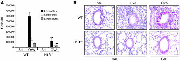
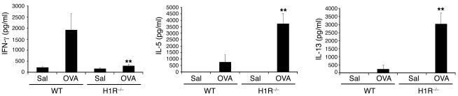
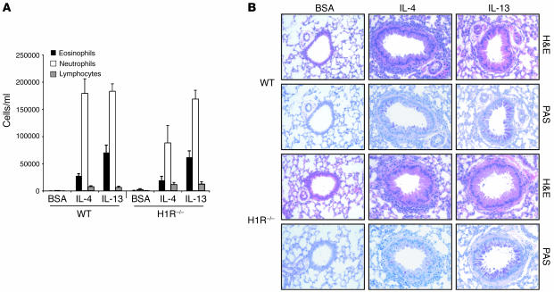
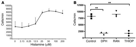
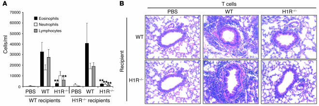
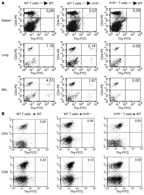
Histamine, signaling via the type 1 receptor (H1R), has been shown to suppress Th2 cytokine production by in vitro cultured T cells. We examined the role of H1R in allergic inflammation in vivo using a murine asthma model. Allergen-stimulated splenic T cells from sensitized H1R-/- mice exhibited enhanced Th2 cytokine production. Despite this Th2 bias, allergen-challenged H1R-/- mice exhibited diminished lung Th2 cytokine mRNA levels, airway inflammation, goblet cell metaplasia, and airway hyperresponsiveness (AHR). Restoration of pulmonary Th2 cytokines in H1R-/- mice by intranasal IL-4 or IL-13 restored inflammatory lung responses and AHR. Further investigation revealed that histamine acts as a T cell chemotactic factor and defective T cell trafficking was responsible for the absence of lung inflammation. Cultured T cells migrated in response to histamine in vitro, but this was ablated by blockade of H1R but not H2R. In vivo, allergen-specific WT but not H1R-/- CD4+ T cells were recruited to the lungs of naive recipients following inhaled allergen challenge. H1R-/- T cells failed to confer airway inflammation or AHR observed after transfer of WT T cells. Our data establish a role for histamine and H1R in promoting the migration of Th2 cells into sites of allergen exposure.


Figures

Figure 1
Figure 1. H1R–/– mice do not develop inflammatory airway responses to antigen.
OVA-immunized (OVA) or sham-immunized (Sal) WT or H1R–/– mice were exposed to OVA by inhalation and studied for inflammatory airway changes. (A) The cellular composition of the BAL fluid was determined. Data represent mean ± SEM. n = 9–10 individuals. **P < 0.01 by Student’s t test. (B) Representative histological sections stained with H&E or PAS at ×200 magnification.
Figure 2
Figure 2. Hyperproduction of Th2 cytokines by splenocytes from H1R–/– mice.
Splenocytes were stimulated for 72 hours in the presence of antigen (OVA). Cytokine secretion was determined by sandwich ELISA. Data represent mean ± SEM from 4 individuals per group. **P < 0.01 by Student’s t test, compared with WT OVA values.
Figure 3
Figure 3. IL-4 and IL-13 induce airway inflammation in H1R–/– mice.
Five micrograms of recombinant mouse IL-4 or IL-13 or 5 μg BSA was administered intranasally for 3 days, and inflammation was determined. Each group consisted of 5 mice. (A) The numbers of eosinophils, neutrophils, and lymphocytes in the BAL fluid. (B) Representative airway histology in WT and H1R–/– lung tissue. Sections were examined at ×200.
Figure 4
Figure 4. OVA-specific T cells migrate toward histamine via an H1R-dependent process.
Chemotaxis to histamine was investigated using the OVA-specific DO11.10 T cell hybridoma. (A) DO11.10 cells migrate in response to histamine. Cells were added to the upper chamber of Transwell chambers, and histamine was added to the lower chamber. After 4 hours, the number of cells in the lower chamber was determined by FACS. (B) Diphenhydramine (DPH), an H1R antagonist; ranitidine (RAN), an H2R antagonist; and thioperamide (THIOP), an H3R/H4R antagonist, were added to assess their effects on T cell chemotaxis induced by histamine (100 μM). Data represent the number of cells moving through Transwell chambers (mean cells ± SEM) from 3 separate experiments. **P < 0.01 by Student’s t test.
Figure 5
Figure 5. Inflammatory airway responses to antigen can be induced by WT T cells but not by H1R–/– T cells.
Antigen-expanded CD4+ T cells from WT or H1R–/– mice were adoptively transferred into naive WT or H1R–/– recipients, and the development of airway inflammation was studied after inhaled OVA challenge. (A) The numbers of eosinophils, neutrophils, and lymphocytes in BAL fluid were determined (n = 9/group). **P < 0.01, Student’s t test. (B) Representative lung histology at ×200 magnification. Sections were stained with H&E.
Figure 6
Figure 6. H1R–/– T cells do not migrate into the lung following antigen challenge.
Thy1 congenic antigen-expanded CD4+ T cells (A) or CD3+ T cells (B) from WT or H1R–/– mice were adoptively transferred into naive WT or H1R–/– recipients. Cells were adoptively transferred into naive recipients and their movement into spleen, whole lung tissue, and BAL fluid studied. Single-cell suspensions were stained using FITC-Thy1.1 or FITC-Thy1.2 (see Methods) and PE-CD4 or PE-CD8 to determine the presence of the transferred cells. The plots shown were obtained by analysis of the pooled cells of 5 animals of each group and represent 3 individual experiments.

References

    1. Corrigan C.J., Kay A.B. CD4 T-lymphocyte activation in acute severe asthma. Relationship to disease severity and atopic status. Am. Rev. Respir. Dis. 1990;141:970–977. - PubMed
    1. Robinson D.S., et al. Predominant TH2-like bronchoalveolar T-lymphocyte population in atopic asthma. N. Engl. J. Med. 1992;326:298–304. - PubMed
    1. Robinson D., et al. Activation of CD4+ T cells, increased TH2-type cytokine mRNA expression, and eosinophil recruitment in bronchoalveolar lavage after allergen inhalation challenge in patients with atopic asthma. J. Allergy Clin. Immunol. 1993;92:313–324. - PubMed
    1. Finkelman F.D., et al. IL-4 is required to generate and sustain in vivo IgE responses. J. Immunol. 1988;141:2335–2341. - PubMed
    1. Lee J.J., et al. Interleukin-5 expression in the lung epithelium of transgenic mice leads to pulmonary changes pathognomonic of asthma. J. Exp. Med. 1997;185:2143–2156. - PMC - PubMed

Publication types

MeSH terms