Comparative Study
. 2006 Jul 1;497(1):101-14.
doi: 10.1002/cne.20985.

Analysis of direct hippocampal cortical field CA1 axonal projections to diencephalon in the rat

Affiliations
Comparative Study

Analysis of direct hippocampal cortical field CA1 axonal projections to diencephalon in the rat

Lee A Cenquizca et al. J Comp Neurol. .

Abstract

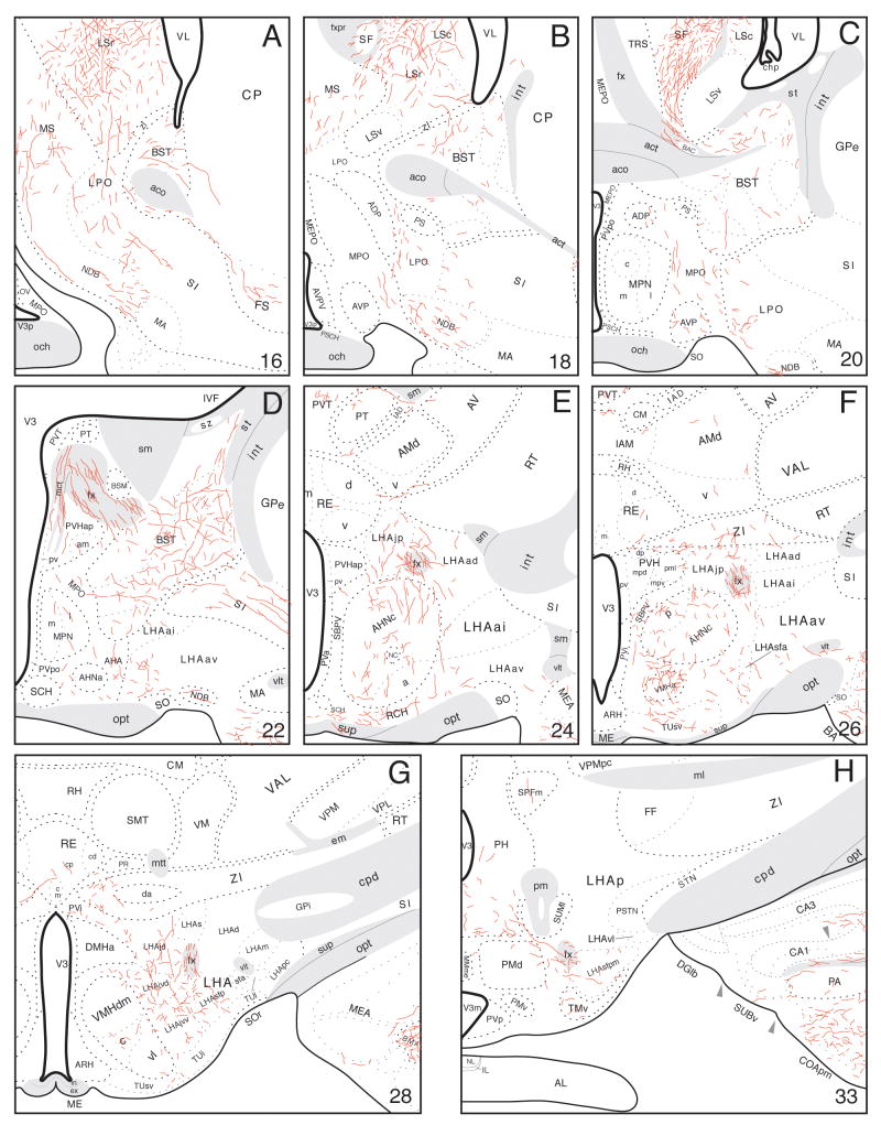
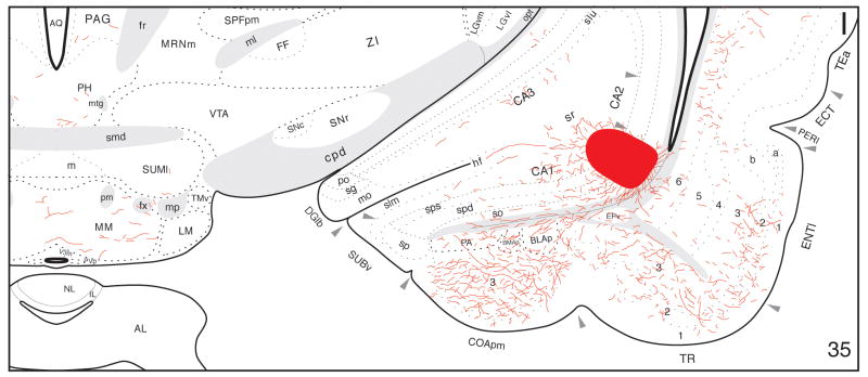
The hippocampal formation is generally considered essential for processing episodic memory. However, the structural organization of hippocampal afferent and efferent axonal connections is still not completely understood, although such information is critical to support functional hypotheses. The full extent of axonal projections from field CA1 to the interbrain (diencephalon) is analyzed here with the Phaseolus vulgaris-leucoagglutinin (PHAL) method. The ventral pole of field CA1 establishes direct pathways to, and terminal fields within, the anterior hypothalamic nucleus, ventromedial hypothalamic nucleus, lateral hypothalamic and lateral preoptic areas, medial preoptic area, and certain other hypothalamic regions, as well as particular midline thalamic nuclei. These results suggest that hippocampal field CA1 modulates motivated or goal-directed behaviors, and physiological responses, associated with the targeted hypothalamic neuron populations.


Figures

Fig. 1
Fig. 1
Distribution of injection sites centered in hippocampal field CA1. Relative locations of PHAL injections from 62 experiments, plotted onto an unfolded map of the rat hippocampal formation (adapted from Petrovich et al., 2001). Injection sites filled with black illustrate experiments with injection sites limited to, or contained within, the ventral pole of field CA1. This triangular region occupies about 10% of field CA1's total surface area.
Fig. 2
Fig. 2
The ventral field CA1 PHAL injection site (black oval regions) in Experiment HIPPO103 (A-C). Panel A is a photomicrograph through the rostral end of the injection site, panel B through the middle, and panel C through the caudal end of the injection site. Panels D-F are photomicrographs of nearby thionin (Nissl)-stained sections to show cytoarchitectonic features of the transverse histological sections (paired with panels A-C, respectively). As currently understood, the major architectonic feature that distinguishes field CA1 from the subiculum is the presence of a clear stratum oriens in the former. See Figures 1 and 3-I for relative location of this injection site. Scale = 200 μm.
Fig. 3
Fig. 3
A-I: Distribution of PHAL-labeled axons (red lines) plotted onto a series of rat brain reference drawings (adapted from Swanson, 2004), cut in the transverse plane and arranged from rostral (A) to caudal (I)—after an injection centered in the ventral pole of hippocampal field CA1 (red area in I).
Fig. 3
Fig. 3
A-I: Distribution of PHAL-labeled axons (red lines) plotted onto a series of rat brain reference drawings (adapted from Swanson, 2004), cut in the transverse plane and arranged from rostral (A) to caudal (I)—after an injection centered in the ventral pole of hippocampal field CA1 (red area in I).
Fig. 4
Fig. 4
A-B: Darkfield photomicrographs showing the distribution of PHAL-labeled axons in the hypothalamus at the level of the anterior hypothalamic nucleus (A), and the ventromedial hypothalamic nucleus (B). C-D: Brightfield photomicrographs demonstrating the location of the injection site (C) and an adjacent thionin-stained section (D) in Experiment HIPPO161. Scale bars: 200 μm.
Fig. 5
Fig. 5
Darkfield photomicrograph showing the divergence of PHAL-labeled axons from the fimbria to the medial corticohypothalamic tract, in a transverse section from an experiment with an injection confined to the ventral pole of hippocampal field CA1. Figure 3D is at approximately the level of this photomicrograph. Scale bar: 200 μm.
Fig. 6
Fig. 6
Summary diagram to indicate the general organization of projections from the ventral pole of hippocampal field CA1 to the interbrain (diencephalon). Projections to other parts of the forebrain are not shown. The flatmap of the rat central nervous system is based on Swanson (2004); line thickness is roughly proportional to relative projection strength.

References

    1. Albers HE, Rawls S. Coordination of hamster lordosis and flank marking behavior: role of arginine vasopressin within the medial preoptic-anterior hypothalamus. Brain Res Bull. 1989;23:105–109. - PubMed
    1. Allen GV, Hopkins DA. Mamillary body in the rat: topography and synaptology of projections from the subicular complex, prefrontal cortex, and midbrain tegmentum. J Comp Neurol. 1989;286:311–336. - PubMed
    1. Anderson P, Bliss TV, Skrede KK. Lamellar organization of hippocampal pathways. Exp Brain Res. 1971;13:571–591. - PubMed
    1. Andrade JP, Madeira MD, Paula-Barbosa MM. Sexual dimorphism in the subiculum of the rat hippocampal formation. Brain Res. 2000;875:125–137. - PubMed
    1. Bamshad M, Albers HE. Neural circuitry controlling vasopressin-stimulated scent marking in Syrian hamsters (Mesocricetus auratus) J Comp Neurol. 1996;369:252–263. - PubMed

Publication types