Comparative Study
. 2006 Jul 1;497(1):1-12.
doi: 10.1002/cne.20954.

Nucleoside triphosphate diphosphohydrolase-2 is the ecto-ATPase of type I cells in taste buds

Affiliations
Comparative Study

Nucleoside triphosphate diphosphohydrolase-2 is the ecto-ATPase of type I cells in taste buds

Dianna L Bartel et al. J Comp Neurol. .

Abstract

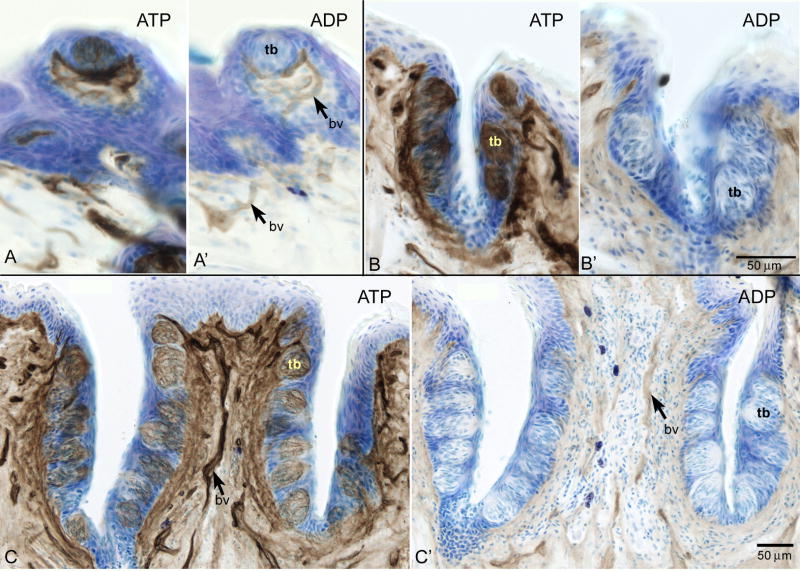
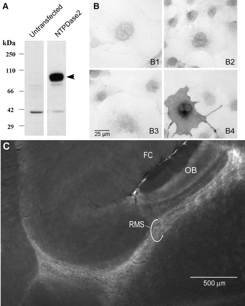
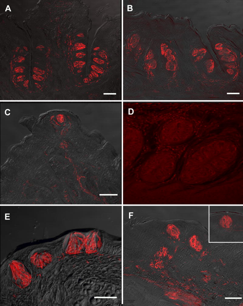
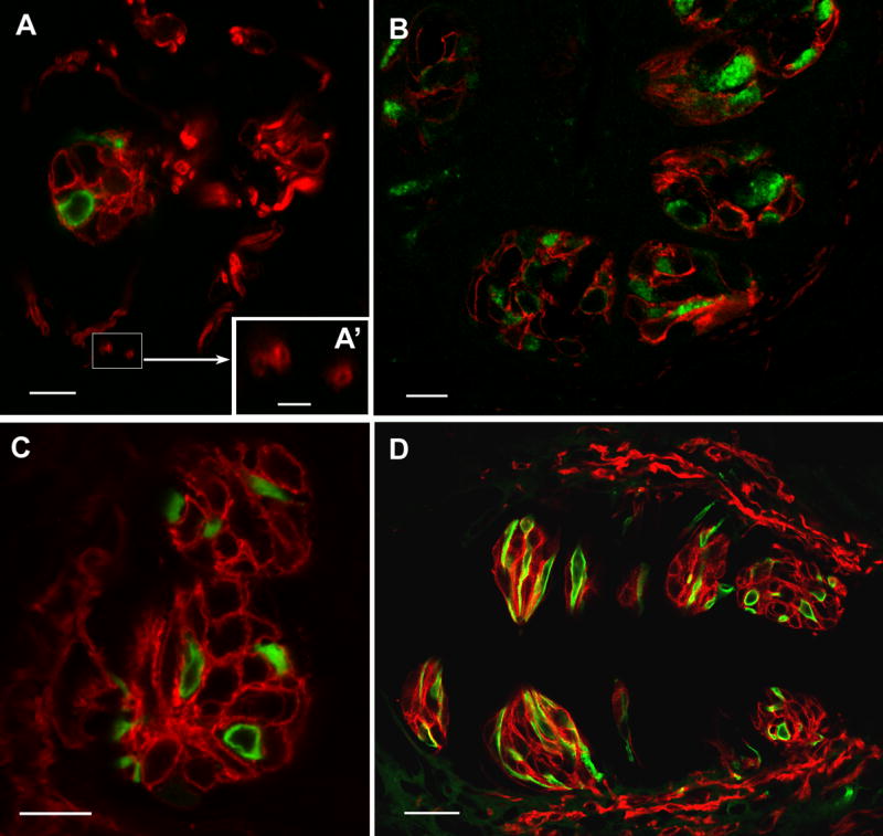
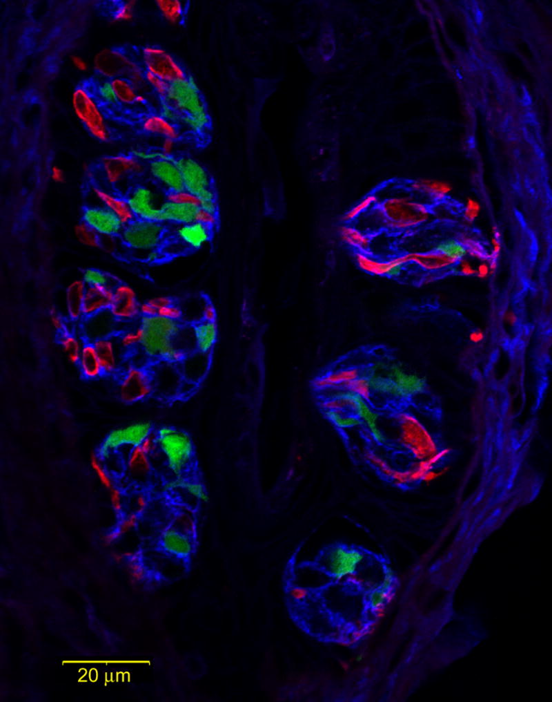
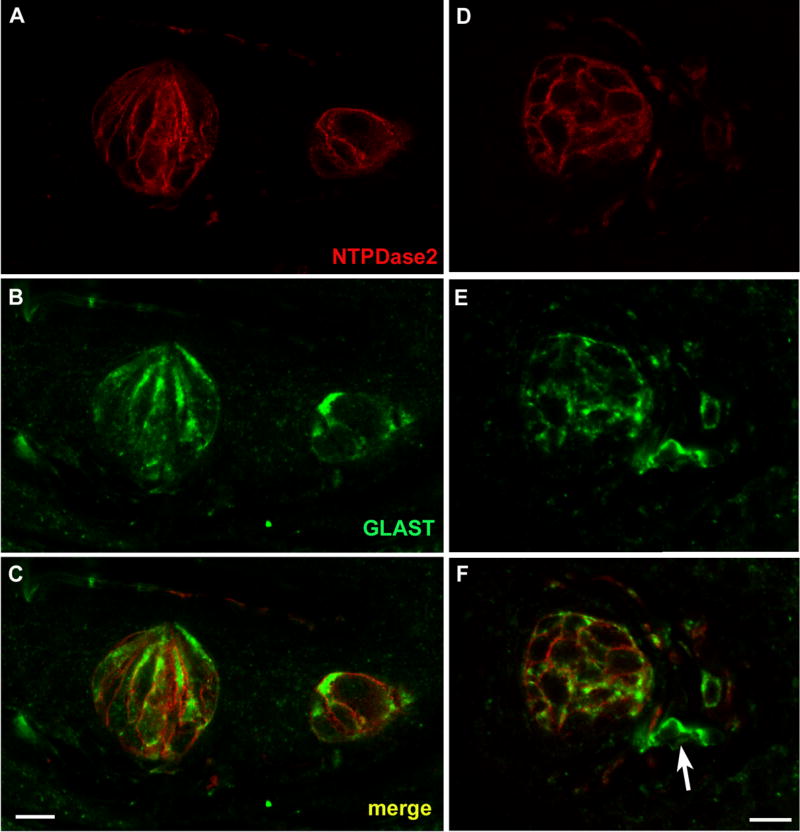
The presence of one or more calcium-dependent ecto-ATPases (enzymes that hydrolyze extracellular 5'-triphosphates) in mammalian taste buds was first shown histochemically. Recent studies have established that dominant ecto-ATPases consist of enzymes now called nucleoside triphosphate diphosphohydrolases (NTPDases). Massively parallel signature sequencing (MPSS) from murine taste epithelium provided molecular evidence suggesting that NTPDase2 is the most likely member present in mouse taste papillae. Immunocytochemical and enzyme histochemical staining verified the presence of NTPDase2 associated with plasma membranes in a large number of cells within all mouse taste buds. To determine which of the three taste cell types expresses this enzyme, double-label assays were performed with antisera directed against the glial glutamate/aspartate transporter (GLAST), the transduction pathway proteins phospholipase Cbeta2 (PLCbeta2) or the G-protein subunit alpha-gustducin, and serotonin (5HT) as markers of type I, II, and III taste cells, respectively. Analysis of the double-labeled sections indicates that NTPDase2 immunoreactivity is found on cell processes that often envelop other taste cells, reminiscent of type I cells. In agreement with this observation, NTPDase2 was located to the same membrane as GLAST, indicating that this enzyme is present in type I cells. The presence of ecto-ATPase in taste buds likely reflects the importance of ATP as an intercellular signaling molecule in this system.


Figures

Figure 1
Figure 1
Massively parallel signature sequencing (MPSS) A: The genomic locations and structures of Entpd1, Entpd2, Entpd3 and Entpd8 (genes encoding NTPDases 1, 2, 3 and 8) are shown. Gene chromosomal positions were obtained from the Build 33 assembly by NCBI. Exons are indicated by filled boxes, and the position and direction of the signature tags are indicated by arrowheads. As expected from the cloning procedures, each signature is associated with the 3′ most DpnII site of the coding sequence. B: Histograms display the abundance of each signature, expressed as transcripts per million (tpm), in libraries constructed from mRNA isolated from either taste bud enriched tissue (TB), lingual epithelium devoid of taste buds (LE), or inner ear derived tissues including the Organ of Corti (Cr), stria vascularis (SV) and otic vesicle (OV). Entpd2 mRNA is represented 9 times per million in the TB library (arrowhead below bar graph) but is not represented in the LE library, suggesting selective Entpd2 expression in taste buds. Taken together with the absence of Entpd1, Entpd3, and Entpd8 in the TB library, these results support that NTPDase2 is the primary ecto-ATPase expressed in taste buds. For sake of comparison, the taste receptor T1R2 is represented 10 tpm in the TB library and is absent in the LE library (data not shown).
Figure 2
Figure 2
Enzyme histochemical analysis revealing ecto-ATPase and ecto-ADPase activity via lead sulfide precipitate (brown) with thionin couterstain (blue) in taste buds in fungiform (A,A’), foliate (B, B’) and circumvallate (C, C’) papillae. With ATP as a substrate (A, B, C) strong ecto-ATPase activity is present in all taste buds (tb). Substituting ADP as a substrate results in lack of activity in the taste buds (A’, B’, C’) though some ecto-ADPase activity is present in nearby blood vessels (bv) (A’, C’). Scale bar in B’ also applies to A, A’, B. Scale bar in C applies to C’.
Figure 3
Figure 3
Anti-NTPDase2 polyclonal antibody specificity A: Western blot analysis of lysate from untransfected COS-7 cells compared to lysate from COS-7 cells transfected with the expression vector plasmid containing the entire NTPDase2 cDNA. As expected, the transfected cells show a prominent band of ~75 kDa (arrowhead) confirming the presence of NTPDase2. B: Immunocytochemistry on intact COS-7 cells transfected (B3,B4) or untransfected (B1,B2) with NTPDase2 cDNA. Cells were incubated with the pre-immune serum (B1,B3) or the anti-NTPDase2 antibody (B2,B4). Immunoreactivity is present only with transfected cells incubated with the NTPDase2 antibody (B4). C: Immunofluorescence for NTPDase2 in a parasagittal brain section (anterior to the right) showing strong expression of NTPDase2 in the rostral migratory stream (RMS) connecting to the olfactory bulb (OB). Staining with this antibody correlates with the previously described distribution of NTPDase2 in the RMS (Braun et al., 2003). FC = frontal cortex.
Figure 4
Figure 4
Nomarski images overlaid with images showing for NTPDase2 in mouse taste buds and surrounding fibers. A: circumvallate papillae, B: foliate papillae, C: fungiform papillae, E: naso-incisor ducts, F: palate, insert: larynx. Incubation with the preimmune serum did not reveal any significant fluorescent signal in the foliate papillae shown in D (taken at eight times the exposure of the other panel figures). Scale bars = 50μm. Scale bar in F also applies to F insert and D.
Figure 5
Figure 5
LSCM images of double label immunocytochemistry with NTPDase2 and the type II cell marker, PLCβ2 or the type III cell marker, 5HT. A, B: Staining for NTPDase2 (red) and PLCβ2 (green) in oblique sections cut through the fungiform (A) and foliate papillae (B). The NTPDase-immunoreactive cells are different from those exhibiting reactivity to either PLCβ2 or 5HT. NTPDase2 reactivity is also associated with nerve processes. Close observation of cross sections of this staining reveals that NTPDase2 immunoreactivity is limited to the periphery of the nerve profile while the core of the fiber remains void of staining, indicative either of membrane-associated neural reactivity or of glial cells circling the fiber (A’, scale bar 2.5μm) C, D: NTPDase2 (red) and 5HT (green) in foliate papillae (C) and cirumvallate papillae (D). Scale bars in A, B and C are 10μm; D scale bar is 25μm.
Figure 6
Figure 6
LSCM images of triple label staining in the foliate papillae with NTPDase2 in blue, the type II cell marker, gustducin in green (GFP), and the type III cell marker, 5HT, in red. NTPDase2-immunoreactive (-ir) cells wrap around the other two cell types, strongly reminiscent of type I cells. NTPDase2-ir type I cells also account for a greater number of cells compared to the other cell types.
Figure 7
Figure 7
LSCM images of double label assays with NTPDase2 and the type I cell marker, GLAST. A, D: NTPDase2 reactive cells in foliate and fungiform papillae, respectively. B, E: GLAST reactive cells in the same sections of foliate and fungiform papillae. C, F: Merged images of NTPDase2 and GLAST revealing colocalization of these markers to the same cellular membranes. The GLAST reactivity extends deeper into the cytoplasm than does the NTPDase2, and so appears as a green halo inside of the double-label (yellow) plasma membrane. GLAST reactivity is also associated with nerve fibers, though not all GLAST positive nerve fibers are double labeled with NTPDase2 as indicated with the arrow in F.

Similar articles

Cited by

References

    1. Abbracchio MP, Burnstock G. Purinergic signaling-pathophysiological roles. Jpn J Pharmacol. 1998;78:113–145. - PubMed
    1. Adler E, Hoon MA, Mueller KL, Chandrashekar J, Ryba NJ, Zuker CS. A novel family of mammalian taste receptors. Cell. 2000;100:693–702. - PubMed
    1. Akisaka T, Oda M. The fine structural localization of adenosine triphosphatase activity on the taste bud in the fungiform papillae of the rat. Arch Histol Jpn. 1977;40:63. - PubMed
    1. Barry M. Ecto-calcium-dependent ATPase activity of mammalian taste buds cells. J Histochem Cytochem. 1992;40:1919–1928. - PubMed
    1. Baryshnikov SG, Rogachevskaja OA, Kolesnikov SS. Calcium signaling mediated by P2Y receptors in mouse taste cells. J Neurophysiol. 2003;90:3283–3294. - PubMed

Publication types

MeSH terms