

Purification and spectroscopic characterization of Ctb, a group III truncated hemoglobin implicated in oxygen metabolism in the food-borne pathogen Campylobacter jejuni
- PMID: 16681372
- PMCID: PMC2528550
- DOI: 10.1021/bi052247k
Purification and spectroscopic characterization of Ctb, a group III truncated hemoglobin implicated in oxygen metabolism in the food-borne pathogen Campylobacter jejuni
Abstract
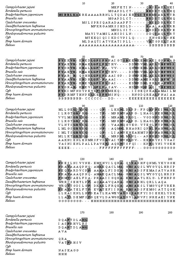
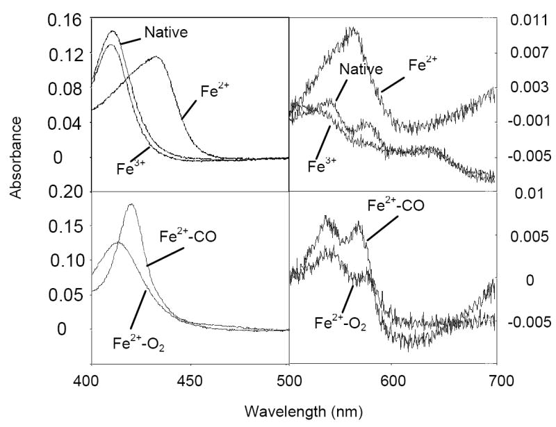
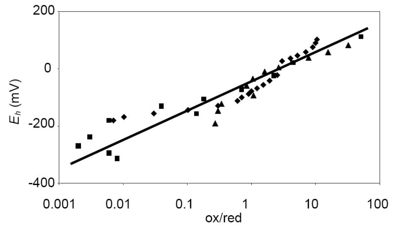
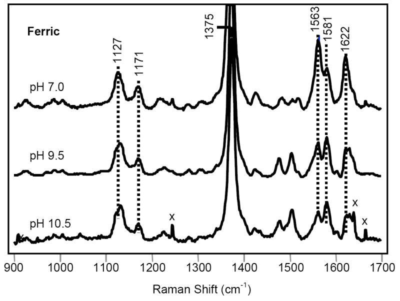
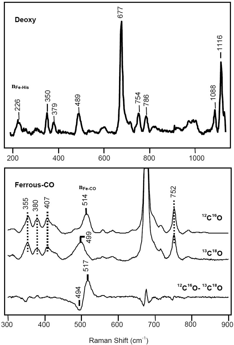
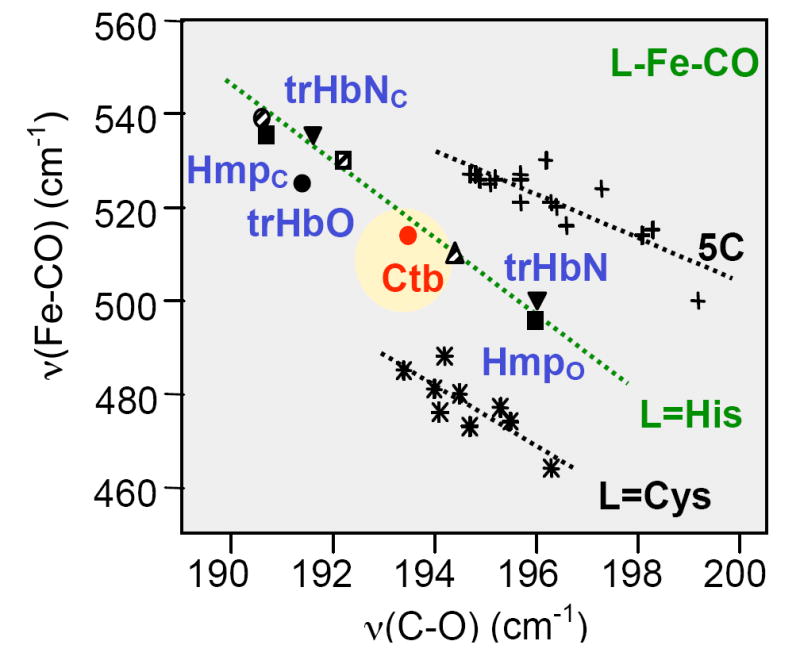
Campylobacter jejuni is a food-borne bacterial pathogen that possesses two distinct hemoglobins, encoded by the ctb and cgb genes. The former codes for a truncated hemoglobin (Ctb) in group III, an assemblage of uncharacterized globins in diverse clinically and technologically significant bacteria. Here, we show that Ctb purifies as a monomeric, predominantly oxygenated species. Optical spectra of ferric, ferrous, O(2)- and CO-bound forms resemble those of other hemoglobins. However, resonance Raman analysis shows Ctb to have an atypical nu(Fe)(-)(CO) stretching mode at 514 cm(-)(1), compared to those of the other truncated hemoglobins that have been characterized so far. This implies unique roles in ligand stabilization for TyrB10, HisE7, and TrpG8, residues highly conserved within group III truncated hemoglobins. Because C. jejuni is a microaerophile, and a ctb mutant exhibits O(2)-dependent growth defects, one of the hypothesized roles of Ctb is in the detoxification, sequestration, or transfer of O(2). The midpoint potential (E(h)) of Ctb was found to be -33 mV, but no evidence was obtained in vitro to support the hypothesis that Ctb is reducible by NADH or NADPH. This truncated hemoglobin may function in the facilitation of O(2) transfer to one of the terminal oxidases of C. jejuni or, instead, facilitate O(2) transfer to Cgb for NO detoxification.
Figures
Similar articles
-
Structural and functional properties of a truncated hemoglobin from a food-borne pathogen Campylobacter jejuni.J Biol Chem. 2007 May 4;282(18):13627-36. doi: 10.1074/jbc.M609397200. Epub 2007 Mar 5. J Biol Chem. 2007. PMID: 17339325
-
Nitric oxide reactivities of the two globins of the foodborne pathogen Campylobacter jejuni: roles in protection from nitrosative stress and analysis of potential reductants.Nitric Oxide. 2013 Nov 1;34:65-75. doi: 10.1016/j.niox.2013.06.002. Epub 2013 Jun 11. Nitric Oxide. 2013. PMID: 23764490
-
The NO-responsive hemoglobins of Campylobacter jejuni: concerted responses of two globins to NO and evidence in vitro for globin regulation by the transcription factor NssR.Nitric Oxide. 2011 Aug 1;25(2):234-41. doi: 10.1016/j.niox.2010.12.009. Epub 2011 Jan 1. Nitric Oxide. 2011. PMID: 21199674
-
The globins of Campylobacter jejuni.Adv Microb Physiol. 2013;63:97-145. doi: 10.1016/B978-0-12-407693-8.00004-2. Adv Microb Physiol. 2013. PMID: 24054796 Review.
-
Hemoglobins from Mycobacterium tuberculosis and Campylobacter jejuni: a comparative study with resonance Raman spectroscopy.Methods Enzymol. 2008;437:255-86. doi: 10.1016/S0076-6879(07)37014-6. Methods Enzymol. 2008. PMID: 18433633 Review.
Cited by
-
Nitrosylation mechanisms of Mycobacterium tuberculosis and Campylobacter jejuni truncated hemoglobins N, O, and P.PLoS One. 2014 Jul 22;9(7):e102811. doi: 10.1371/journal.pone.0102811. eCollection 2014. PLoS One. 2014. PMID: 25051055 Free PMC article.
-
Nitrosative stress defences of the enterohepatic pathogenic bacterium Helicobacter pullorum.Sci Rep. 2017 Aug 30;7(1):9909. doi: 10.1038/s41598-017-10375-1. Sci Rep. 2017. PMID: 28855660 Free PMC article.
-
Recombinant Escherichia coli strains with inducible Campylobacter jejuni single domain hemoglobin CHb expression exhibited improved cell growth in bioreactor culture.PLoS One. 2015 Mar 6;10(3):e0116503. doi: 10.1371/journal.pone.0116503. eCollection 2015. PLoS One. 2015. PMID: 25748170 Free PMC article.
-
Oxygen- and NssR-dependent globin expression and enhanced iron acquisition in the response of campylobacter to nitrosative stress.J Biol Chem. 2008 Oct 17;283(42):28413-25. doi: 10.1074/jbc.M801016200. Epub 2008 Aug 5. J Biol Chem. 2008. PMID: 18682395 Free PMC article.
-
Antimicrobial Activity of the Manganese Photoactivated Carbon Monoxide-Releasing Molecule [Mn(CO)3(tpa-κ(3)N)](+) Against a Pathogenic Escherichia coli that Causes Urinary Infections.Antioxid Redox Signal. 2016 May 10;24(14):765-80. doi: 10.1089/ars.2015.6484. Epub 2016 Mar 30. Antioxid Redox Signal. 2016. PMID: 26842766 Free PMC article.
References
-
- Wu G, Wainwright LM, Poole RK. Microbial globins. Adv Microb Physiol. 2005;47:255–310. - PubMed
-
- Membrillo-Hernández J, Coopamah MD, Anjum MF, Stevanin TM, Kelly A, Hughes MN, Poole RK. The flavohemoglobin of Escherichia coli confers resistance to a nitrosating agent, a “nitric oxide releaser,” and paraquat and is essential for transcriptional responses to oxidative stress. J Biol Chem. 1999;274:748–54. - PubMed
-
- Crawford MJ, Goldberg DE. Role for the Salmonella flavohemoglobin in protection from nitric oxide. J Biol Chem. 1998;273:12543–7. - PubMed
Publication types
MeSH terms
Substances
Grants and funding
LinkOut - more resources
Full Text Sources
Molecular Biology Databases
Research Materials
Miscellaneous
