. 2006 May 16;45(19):6095-104.
doi: 10.1021/bi060138f.

cAMP-Coupled riboflavin trafficking in placental trophoblasts: a dynamic and ordered process

Affiliations

cAMP-Coupled riboflavin trafficking in placental trophoblasts: a dynamic and ordered process

Vanessa M D'Souza et al. Biochemistry. .

Abstract

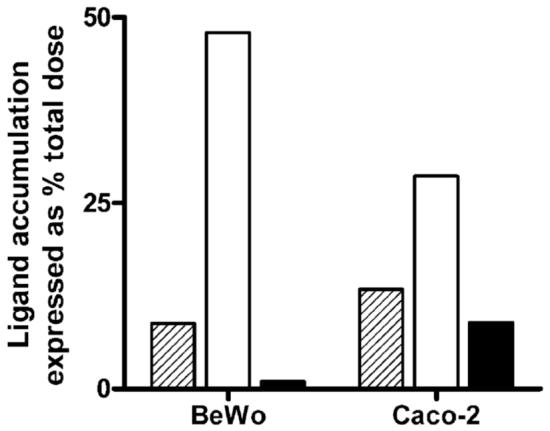
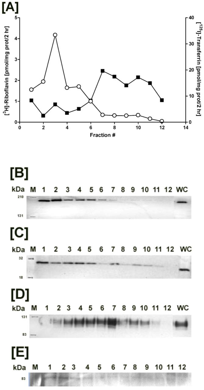
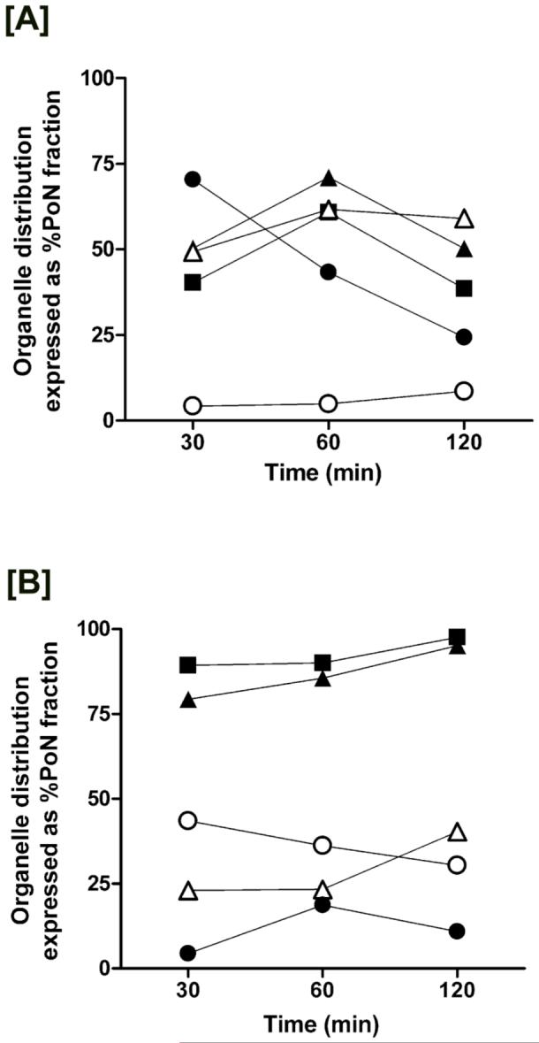
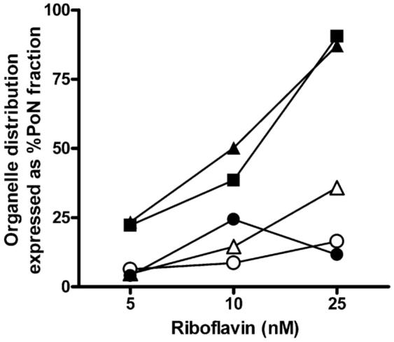
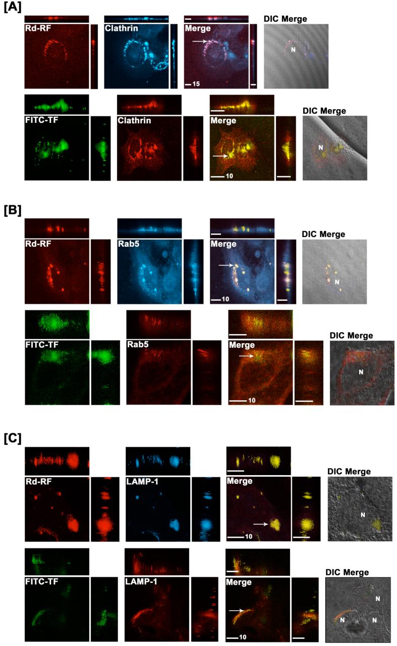
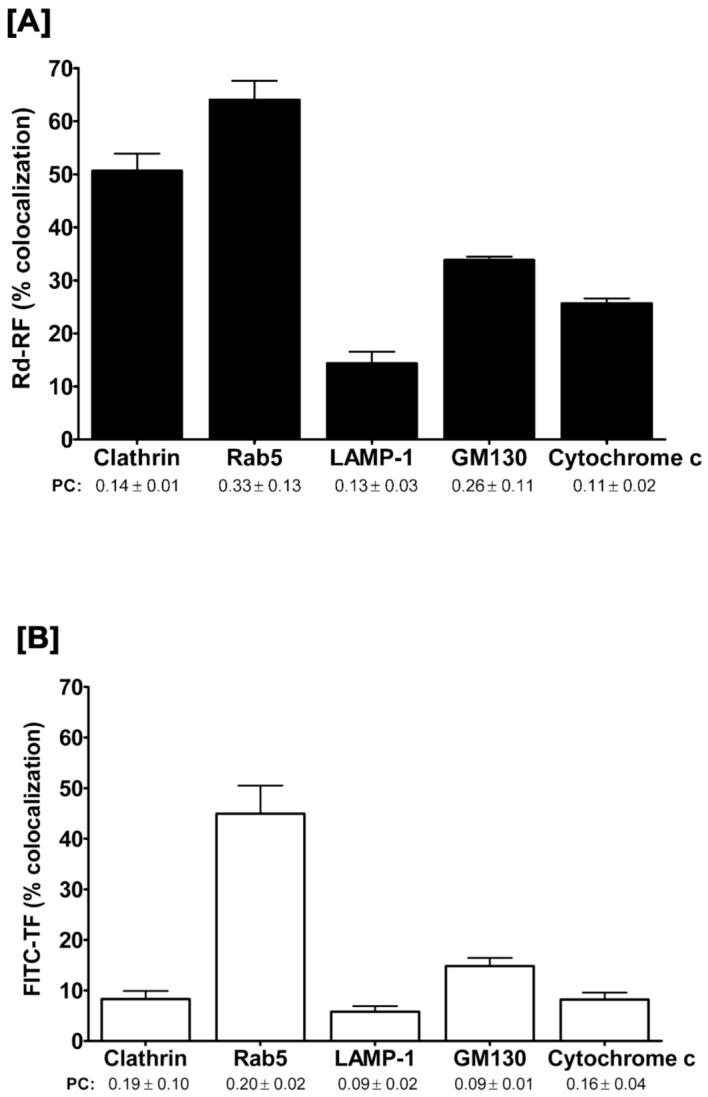
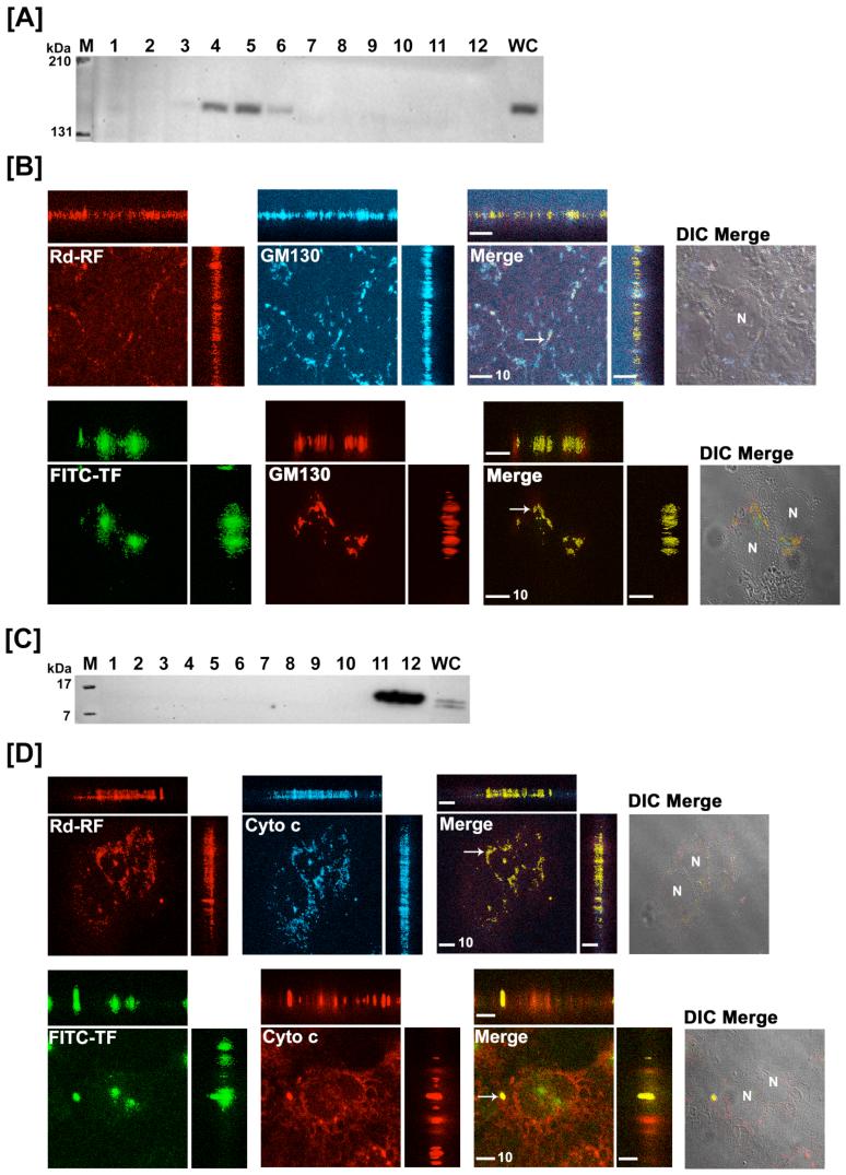
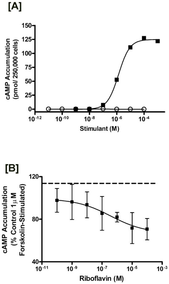
Riboflavin (RF, vitamin B(2)), an essential micronutrient central to cellular metabolism through formation of flavin mononucleotide (FMN) and flavin adenine dinucleotide (FAD) cofactors, is internalized, at least in part, via a proposed receptor-mediated endocytic (RME) process. The purpose of this study was to delineate the cellular RF distribution using human placental trophoblasts and evaluate the regulatory role of cAMP in this process. Subcellular fractionation and three-dimensional confocal microscopy analyses were carried out to define the RF accumulation profile. Biochemical assays evaluating the cAMP dependence of this pathway were also performed. This study records an intracellular RF distribution pattern that shows dynamic accumulation of the ligand predominantly in the endosomal and lysosomal compartments and to a lesser extent in the Golgi and mitochondria. In contrast, transferrin (TF) colocalizes rapidly within endosomes with minimal accumulation in the other organelles. The temporal and spatial distribution of RF and TF colocalized with unique markers of the endocytic machinery provides added morphological evidence in support of the RME process with ultimate translocation to the mitochondrial domain. Colocalized staining with the Golgi also suggests a possible recycling or exocytic mechanism for this ligand. Furthermore, this study demonstrates cAMP regulation of the putative ligand-bound RF receptor and its association into endocytic vesicles. Delineating the dynamics of the process governing cellular RF homeostasis presents an untapped resource that can be further exploited in improving our current understanding of nutritional biology and fetal growth and development, and perhaps in targeting the endogenous system for developing novel therapeutic approaches.


Figures

Figure 1
Figure 1
Comparative cell-associated profiles of [3H]-Riboflavin (10 nM RF) with the endocytic marker, 125I-Transferrin (10 nM TF), and an unrelated bile acid transporter substrate, [3H]-Taurocholic acid (20 nM TCA) in placental (BeWo) and intestinal (Caco-2) cells. Ligand content derived from nuclear and postnuclear fractions and expressed as % total dose revealed ≥ 8% RF accumulation (hashed bars) while TF levels (open bars) varied from 48% in BeWo cells to 28% in Caco-2 cells. Distribution profile of TCA (solid bars) showed negligible accumulation in trophoblasts as compared to enterocytes.
Figure 2
Figure 2
Distribution of [3H]-Riboflavin (RF) and 125I-Transferrin (TF) in the postnuclear fractions isolated from trophoblasts. [A] Postnuclear fractions isolated from BeWo cells were resolved based on differential organelle densities using a discontinuous sucrose gradient, fractionated, and measured for dual radiolabel accumulation (closed squares - [3H]-RF; open circles - 125I-TF). Organelle-enriched fractions were identified by western blot analyses using antibodies directed against endosomal proteins namely clathrin [B] and Rab5 GTPase [C], and the lysosomal marker LAMP1 [D]. [E] Intact radiolabel on TF following homogenization and gradient fractionation was visualized at ∼ 83 kDa by gel electrophoresis and autoradiographic exposure of the isolated fractions.
Figure 3
Figure 3
Time-dependent localization of [3H]-RF and 125I-TF within the separated fractions from BeWo cells. Sucrose density gradient fraction analysis by western blotting of organelle-associated markers (closed squares - clathrin; closed triangles - Rab5 GTPase; open triangles - LAMP1; open circles - GM130; closed circles - cytochrome c) following 30 min, 60 min, and 120 min exposure to RF [A] and TF [B] revealed ligand-specific distribution profiles.
Figure 4
Figure 4
Concentration-dependent trafficking profiles of [3H]-RF after 2 hr incubation at 37 °C. Ligand accumulation of 5 - 25 nM RF in endosomes (closed squares - clathrin; closed triangles - Rab5 GTPase), lysosomes (open triangles - LAMP1), Golgi (open circles - GM130), and mitochondria (closed circles - cytochrome c) increased in a dose-dependent manner in placental trophoblasts.
Figure 5
Figure 5
Endosomal and lysosomal colocalization of Rhodamine-RF (Rd-RF) and FITC-labeled transferrin (FITC-TF). Rd-RF or FITC-TF were examined for colocalization with immunostained early endocytic (clathrin [A] and Rab5 GTPase, [B]), and lysosomal (LAMP1, [C]) protein markers after 60 minutes of ligand internalization in BeWo cells. Images represent orthogonal 3-D profiles with the inset view defining the XY axis and the outer panels reveal the YZ (right narrow panel) and XZ (upper narrow panel) focal planes. Fluorescence signals for each channel were merged to reveal regions of colocalization (indicated by arrows and yellow regions) and overlayed with the corresponding differential interference contrast (DIC) image to define cell morphology (far right inset view). Both Rd-RF and FITC-TF exhibited distinct colocalization with early endosome markers for clathrin and Rab5 GTPase. In contrast to FITC-TF, Rd-RF showed extensive colocalized signal with LAMP1. The nucleus is represented by ‘N’. Scale bars (μm) are defined in the merged inset views.
Figure 6
Figure 6
Quantitative evaluation of the 3-D colocalized regions of RF and TF, respectively with organelle protein markers. Overlapping, i.e., colocalized, volumes (cm3) for either Rd-RF [A] or FITC-TF [B] with organelle channels were expressed as a percentage of the total volume for Rd-RF or FITC-TF. The Pearson’s Correlation (PC) was chosen to define the similarity of 3-D shapes between ligands and overlapping channels with organelle markers. Data are expressed as the mean ± SEM for 3 - 4 regions of interest. All values were > 0.0 and, thus revealed a positive correlation for pattern recognition between channels.
Figure 7
Figure 7
Translocation of [3H]-RF and [125I]-TF to the Golgi and mitochondria in BeWo cells. Post-nuclear fractions collected after 2 hr internalization of [3H]-RF and [125I]-TF in BeWo cells were immunoblotted for Golgi (GM130, [A]) and mitochondria (cytochrome c, [C]). Confocal 3-D images of either Rd-RF or FITC-TF after 60 minutes internalization in BeWo cells were analyzed for colocalization (arrows and yellow regions) with Golgi [B] and mitochondria [D]. Confocal images are defined by orthogonal profiles as described in figure 5 and fluorescent signals were merged with DIC images (right columns, [B] and [D]). Scale bars represent 10 μm and nuclei (N) are defined in DIC images.
Figure 8
Figure 8
Cyclic AMP regulation of the RF internalization process. [A] Determination of cAMP accumulation in BeWo cells after stimulation with riboflavin or forskolin. BeWo cells cultured as described in the experimental section were stimulated via incubation with the indicated concentration of either forskolin (closed squares) for 10 minutes, or riboflavin (open circles) for 40 minutes. Data are a representative curve from three independent experiments showing similar results. [B] Inhibitory effects of riboflavin on forskolin-stimulated cAMP accumulation. BeWo cells were washed and then incubated with the indicated concentrations of riboflavin dissolved in HF12K (20 mM HEPES buffered F12K, pH 7.4, 37 °C). After 10 minutes HF12K was removed and cells were stimulated with 1μM forskolin in the continued presence of riboflavin. cAMP accumulation was then determined as described in the experimental section. Dashed line indicates 1μM forskolin stimulated accumulation in the absence of riboflavin. Results are expressed as mean S.D. (n = 5).

References

    1. Cooperman JM, Lopez R. Handbook of vitamins. Marcel Dekker; New York: 1991. Riboflavin; pp. 283–310.
    1. Powers HJ. Riboflavin (vitamin B-2) and health. Am. J. Clin. Nutr. 2003;77:1352–60. - PubMed
    1. Huang SN, Swaan PW. Involvement of a receptor-mediated component in cellular translocation of riboflavin. J. Pharmacol. Exp. Ther. 2000;294:117–25. - PubMed
    1. Huang SN, Swaan PW. Riboflavin uptake in human trophoblast-derived BeWo cell monolayers: cellular translocation and regulatory mechanisms. J. Pharmacol. Exp. Ther. 2001;298:264–71. - PubMed
    1. Foraker AB, Khantwal CM, Swaan PW. Current perspectives on the cellular uptake and trafficking of riboflavin. Adv. Drug Deliv. Rev. 2003;55:1467–83. - PubMed

Publication types