. 2006 Jun;8(6):961-71.
doi: 10.1111/j.1462-5822.2005.00676.x.

Niche-specific regulation of central metabolic pathways in a fungal pathogen

Affiliations

Niche-specific regulation of central metabolic pathways in a fungal pathogen

Caroline J Barelle et al. Cell Microbiol. 2006 Jun.

Abstract

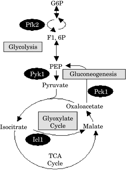
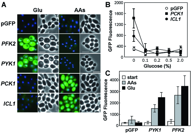
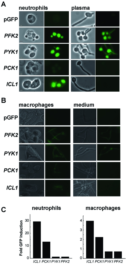
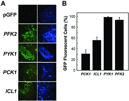
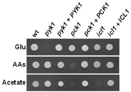
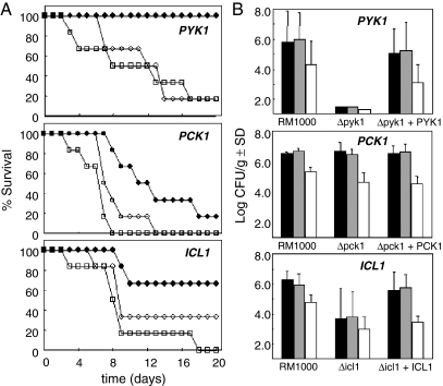
To establish an infection, the pathogen Candida albicans must assimilate carbon and grow in its mammalian host. This fungus assimilates six-carbon compounds via the glycolytic pathway, and two-carbon compounds via the glyoxylate cycle and gluconeogenesis. We address a paradox regarding the roles of these central metabolic pathways in C. albicans pathogenesis: the glyoxylate cycle is apparently required for virulence although glyoxylate cycle genes are repressed by glucose at concentrations present in the bloodstream. Using GFP fusions, we confirm that glyoxylate cycle and gluconeogenic genes in C. albicans are repressed by physiologically relevant concentrations of glucose, and show that these genes are inactive in the majority of fungal cells infecting the mouse kidney. However, these pathways are induced following phagocytosis by macrophages or neutrophils. In contrast, glycolytic genes are not induced following phagocytosis and are expressed in infected kidney. Mutations in all three pathways attenuate the virulence of this fungus, highlighting the importance of central carbon metabolism for the establishment of C. albicans infections. We conclude that C. albicans displays a metabolic program whereby the glyoxylate cycle and gluconeogenesis are activated early, when the pathogen is phagocytosed by host cells, while the subsequent progression of systemic disease is dependent upon glycolysis.


Figures

Fig. 1
Fig. 1
Cartoon of central carbon metabolism, highlighting the steps analysed in this study.
Fig. 2
Fig. 2
Differential in vitro regulation of glycolysis, gluconeogenesis and the glyoxylate cycle in C. albicans.A. C. albicans strains containing PFK2-, PYK1-, PCK1- and ICL1-GFP promoter fusions or the empty pGFP control (CJB-1, CJB-2, CJB-3, CLM1-1, CLM3-2: Table 1) were examined after growth overnight on minimal media containing 2% glucose (Glu) or 2% casamino acids (AAs) as sole carbon source. Merged GFP and DAPI images are shown alongside the corresponding light micrographs. Scale bar represents 10 µm.B. Repression of PCK1- and ICL1-GFP by different concentrations of glucose (mean GFP fluorescence intensity per cell).C. Quantification of mean GFP fluorescence levels for C. albicans cells with PFK2- and PYK1 -GFP fusions resuming growth on glucose or amino acids: cells grown overnight on amino acids (start); cells grown for 2 h on glucose (Glu) or amino acids (AAs).
Fig. 3
Fig. 3
Differential regulation of PFK2-, PYK1-, PCK1- and ICL1-GFP fusions in C. albicans following phagocytosis by (A)neutrophils or (B) macrophages. A. C. albicans cells were mixed with primary human neutrophils in a 1:1 ratio, and examined microscopically after 1.5 h. The ratio of GFP expression in phagocytosed to non-phagocytosed C. albicans cells was measured (C). Corresponding light and fluorescence micrographs of phagocytosed C. albicans cells (neutrophils), and light and fluorescence micrographs of control cells in plasma alone (plasma) are shown: bars represent 10 µm. Fold induction was measured by comparing the mean fluorescence intensity for phagocytosed cells with that for non-phagocytosed C. albicans cells (n > 50). C. albicans strains containing PFK2-, PYK1-, PCK1- or ICL1-GFP fusions were compared with the control carrying the empty vector, pGFP (CJB-1, CJB-2, CJB-3, CLM1-1, CLM3-2: Table 1). B. Cultured murine J774A-1 macrophages were mixed with C. albicans cells in a 1:1 ratio and analysed after 3 h, as described in A: phagocytosed C. albicans cells (macrophages); non-phagocytosed cells (medium). Scale bar represents 20 µm. C. Fold induction of GFP fluorescence in phagocytosed cells versus non-phagocytosed cells in neutrophils and macrophages respectively.
Fig. 4
Fig. 4
Differential regulation of PFK2-, PYK1-, PCK1- and ICL1-GFP fusions in C. albicans in the kidney during systemic candidiasis. A. Corresponding GFP (green) and Calcofluor White-stained (blue) images of C. albicans cells infecting the mouse kidney. C. albicans strains containing PFK2-, PYK1-, PCK1- or ICL1-GFP fusions were compared with the control strain carrying the empty vector, pGFP (CJB-1, CJB-2, CJB-3, CLM1-1, CLM3-2: Table 1). B. Proportion of C. albicans cells (n > 1000) infecting the kidney that display GFP fluorescence above background levels, in animals displaying clinical signs of infection.
Fig. 5
Fig. 5
Growth of C. albicans pyk1/pyk1, pck1/pck1 and icl1/icl1 null mutants on different carbon sources. C. albicans wild type, pyk1/pyk1, pck1/pck1 and icl1/icl1 strains (wt, CLM19-3; pyk1, CLM44-5; pck1, CLM56-4; icl1, CLM25-5: Table 1) and the corresponding control strains containing a reintegrated copy of the wild-type gene (pyk1 + PYK1, CLM45-9; pck1 + PCK1, CLM57-2; icl1 + ICL1, CLM26-1: Table 1) were grown on minimal medium containing 2% glucose (Glu), 2% amino acids (AAs) or 2% acetate as sole carbon source.
Fig. 6
Fig. 6
Virulence of C. albicans pyk1/pyk1, pck1/pck1 and icl1/icl1 null mutants in the mouse model of systemic infection. A. Survival of immunocompetent female BALB/c mice following tail vein injection of C. albicans cells: open squares, first control representing the parental strain, RM1000 containing CIp20 (CLM19-3: Table 1); closed circles, experiment representing the homozygous null mutant containing CIp20 (icl1/icl1, CLM25-5; pck1/pck1, CLM56-4; pyk1/pyk1, CLM44-5: Table 1); open circles, second control representing the null mutant containing CIp20 and the wild-type gene (ICL1, CLM26-1; PCK1, CLM57-2; PYK1, CLM45-9: Table 1). B. Tissue burdens in mouse organs infected with the same C. albicans strains. Fungal colony forming units per gram of tissue were measured in the left kidney (black), right kidney (grey) and brain (white).

Similar articles

Cited by

References

    1. Abi-Said D, Anaissie E, Uzun O, Raad I, Pinzcowski H, Vartivarian S. The epidemiology of hematogenous candidiasis caused by different Candida species. Clin Infect Dis. 1997;24:1122–1128. - PubMed
    1. Barelle CJ, Manson C, MacCallum D, Odds FC, Gow NAR, Brown AJP. GFP as a quantitative reporter of gene regulation in Candida albicans. Yeast. 2004;21:333–340. - PubMed
    1. Brand A, MacCallum DM, Brown AJP, Gow NAR, Odds FC. Ectopic expression of URA3 can influence the virulence phenotypes and proteome of Candida albicans but can be overcome by targeted reintegration of URA3 at the RPS10 locus. Eukaryot Cell. 2004;3:900–909. - PMC - PubMed
    1. Brown AJP. Integration of metabolism with virulence in Candida albicans. In: Brown AJP, editor. Fungal Genomics. Heidelberg: Mycota XIII, Springer-Verlag; 2005. pp. 185–203.
    1. Calderone RA. Candida and Candidiasis. Washington DC: American Society for Microbiology Press; 2002.

Publication types

MeSH terms

LinkOut - more resources