. 2006 May 23;16(10):1041-9.
doi: 10.1016/j.cub.2006.04.005. Epub 2006 May 11.

The miRNA-processing enzyme dicer is essential for the morphogenesis and maintenance of hair follicles

Affiliations

The miRNA-processing enzyme dicer is essential for the morphogenesis and maintenance of hair follicles

Thomas Andl et al. Curr Biol. .

Abstract

The discovery that microRNAs (miRNAs) play important roles in regulating gene expression via posttranscriptional repression has revealed a previously unsuspected mechanism controlling development and progenitor-cell function (reviewed in ); however, little is known of miRNA functions in mammalian organogenesis. Processing of miRNAs and their assembly into the RNA-induced silencing (RISC) complex requires the essential multifunctional enzyme Dicer . We found that Dicer mRNA and multiple miRNAs are expressed in mouse skin, suggesting roles in skin- and hair-follicle biology. In newborn mice carrying an epidermal-specific Dicer deletion, hair follicles were stunted and hypoproliferative. Hair-shaft and inner-root-sheath differentiation was initiated, but the mutant hair follicles were misoriented and expression of the key signaling molecules Shh and Notch1 was lost by postnatal day 7. At this stage, hair-follicle dermal papillae were observed to evaginate, forming highly unusual structures within the basal epidermis. Normal hair shafts were not produced in the Dicer mutant, and the follicles lacked stem cell markers and degenerated. In contrast to decreased follicular proliferation, the epidermis became hyperproliferative. These results reveal critical roles for Dicer in the skin and implicate miRNAs in key aspects of epidermal and hair-follicle development and function.


Figures

Figure 1
Figure 1. Expression of Dicer and miRNAs in the Skin and Generation of an Epidermal-Specific Dicer Deletion
(A) E14.5 mouse embryo subjected to whole-mount in situ hybridization with Dicer probe. Generalized expression is detected in the epidermis (blue staining) with elevated signal in developing hair and whisker follicles (examples indicated by arrows). (B) Section in situ hybridization of P7 dorsal mouse skin with Dicer probe. Intense signal (purple-brown) is detected in the epidermis and hair-follicle outer root sheath (arrows). (C) Semiquantitative RT-PCR analysis of expression of miRNAs mmu-mir-200b, mmu-mir-196a, mmu-mir-29b, and mmu-mir-24 in mouse skin with primers specific for the mature miRNAs. The samples used were as follows: P6, isolated P6 epidermis; P16 ana, P16 posterior dorsal epidermis containing anagen-stage hair follicles; P16 Cat, P16 anterior dorsal epidermis containing catagen-stage hair follicles; P20, anterior dorsal P20 epidermis containing telogen-stage hair follicles; P1C, full-thickness P1 control dorsal skin; and P1 Dkk1, full-thickness dorsal skin from a P1 mouse ectopically expressing Dkk1 in the epidermis and lacking hair follicles. (D) Schematic depiction of PCR primers used for detection of wild-type, floxed, and Cre-excised Dicer alleles. (E) Analysis of dermal and epidermal genomic DNA isolated from newborn K14-Cre; Dicerflox/+, Dicerflox/+, and K14-Cre; Dicerflox/flox mice. The Dicerflox/+ allele is efficiently recombined in the epidermis, but not the dermis of mice carrying K14-Cre. (F) Western blots of isolated dorsal epidermis at P1 (left panel) and full-thickness dorsal skin at P63 (right panel) from littermate control (cont) or Dicer mutant (KO) mice incubated with anti-Dicer antibody (upper panels), anti-p27 (bottom left), or anti-Ezrin (bottom right). Dicer protein is absent from KO epidermis at P1 and greatly reduced in full-thickness skin at P63. (G) Northern blot of RNA from control and Dicer mutant epidermis at P1 (left panel) and P6 (right panel) hybridized with probe for mmu-mir-200b. miRNA precursors are detected in all lanes; the mature processed miRNA is detected in the control lanes but is absent from Dicer mutant epidermis. (H) Semiquantitative RT-PCR analysis of expression of mmu-mir-27b, mmu-mir-203, mmu-mir-24, and mmu-mir-200b in control littermate and Dicer mutant skin with primers specific for the mature miRNAs. P6 RNAs were isolated from epidermis and P1 and P63 RNAs from full-thickness dorsal skin. (I and J) Phenotypes of Dicer mutant mice with control littermates at P7 (I) and P13 (J). Note the complete lack of external hair in the Dicer mutant depicted at P7 (I). The mutant depicted at P13 showed a mosaic phenotype with loss of external hair over the majority of the body, but sparing the head and right flank. (K–M) Whole mounts of dorsal skin from control littermate (K) or Dicer mutant (L and M) at P6 viewed from the epidermal (K and L) or dermal (M) side. Whole-mounted skin was overstained with alkaline phosphatase to reveal skin structure. Note epidermal evaginations in (L) (arrows) and failure of mutant hair shafts to penetrate the epidermis. Mutant hair follicles are misangled (M). (N–V′) Histological analysis of Dicer mutant (O, O′, Q, Q′, S, T, V, and V′) and littermate control (N, N′, P, P′, R, U, and U′) skin from mice at P1 (N–O′), P7 (P–T), and P49 (U–V′). Paraffin sections were stained with hematoxylin and eosin. Arrow in (S) indicates abnormal stunted hair follicle in Dicer mutant skin at P7. Arrows in (T) indicate abnormal evaginations of dermal cells into Dicer null epidermis at P7. Panels (N), (O), (P), (Q), (U), and (V) were photographed at 10× magnification; (P′) and (Q′) at 20×; and (N′), (O′), (R)–(T), (U′), and (V′) at 40×. Size bar in panel (V′) indicates the following: 200 μm for panels (N), (O), (P), (Q), (U), and (V); 100 μm for panels (P′) and (Q′); and 50 μm for panels (N′), (O′), (R)–(T), (U′), and (V′).
Figure 2
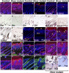
Figure 2. Expression of K15 but Not Shh or Notch1 Is Affected by Loss of Epidermal Dicer in Newborn Skin
(A–F) Immunofluorescence of paraffin sections from newborn Dicer mutant (B, D, and F) and littermate control (A, C, and E) skin. (A) and (B) show immunofluorescence for the suprabasal epidermal marker K10 (green) and the proliferation-associated antigen Ki67 (red). Expression of K10 was similar in the mutant and control, but fewer Ki67-positive cells were detected in mutant hair follicles. (C and D) Immunofluorescence for the proliferation marker phosphohistone H3 (red) indicates that there are fewer proliferating cells in Dicer mutant than control hair follicles (arrows). (E and F) Immunofluorescence for K15 (red) reveals expression in control ([E], arrow) but not Dicer mutant (F) epidermis. Nuclei in panels (A)–(F) are counterstained with DAPI (blue). (G–J) In situ hybridization of paraffin sections from newborn Dicer mutant (H and J) and littermate control (G and I) skin with probes for Shh (G and H) and Notch1 (I and J). Shh is expressed similarly in control and Dicer newborn hair follicles (arrows in [G] and [H]). Notch1 is expressed similarly in control and Dicer newborn hair follicles and epidermis (arrows in [I] and [J]). Panels [A]–[J] were photographed at the same magnification. (K and L) TEM of newborn control (K) and Dicer mutant (L) skin showing apoptotic cell (arrow) next to the basement membrane (BM) in the mutant. (M) Representative immunoblots of P1 control littermate and Dicer mutant epidermis incubated with antibodies to the indicated proteins. Dicer protein is absent in mutant epidermis. Expression of phosphohistone H3 (P-H3) and SOX9 is reduced and the cleaved form of caspase 3 is markedly elevated in the mutant.
Figure 3
Figure 3. Dicer Mutant Skin at P7 Is Characterized by Lack of Shh and Notch1 Expression, Proliferative Defects, and Evagination of Dermal Cells into the Epidermis
(A–F) Paraffin sections of skin from P7 Dicer mutant (B, D, and F) and littermate control (A, C, and E) subjected to immunofluorescence (red) for K14 (A and B), K1 (C and D), and S100A3 (E and F). Basal and suprabasal epidermal cell layers are expanded in the Dicer mutant. Differentiating hair-shaft cells are present in Dicer mutant hair follicles, but at lower numbers than in the control. (G–N) Paraffin sections of skin from P7 Dicer mutant (H, J, L, and N) and littermate control (G, I, K, and M) subjected to in situ hybridization with the digoxygenin-labeled probes indicated. Positive signals appear purple brown and are indicated by arrows. Pigmented cells of the hair shaft appear black. Hybridization for Shh, Gli1, and Crabp1 was absent in Dicer mutant hair follicles, and Notch1 expression was absent from the mutant epidermis and severely reduced in hair follicles. (O–P′) Immunofluorescence detection of BrdU in Dicer mutant (P and P′) and littermate control (O and O′) skin at P7. Labeled cells appear red. Their numbers are decreased in mutant hair follicles but increased in the epidermis compared with the control (arrows). (Q and R) P7 Dicer mutant (R) and control (Q) skin subjected to TUNEL staining (green) and immunofluorescence for K17 (red). Note expanded K17 staining in the mutant epidermis. The frequency of TUNEL-positive cells is increased in Dicer mutant epidermis and hair follicles (arrows in [R]). (S–X′) Immunoflourescence detection of GATA3 (S and T) (red), SOX9 (U and V) (red), K14 (W and X) (red), and K17 (W′ and X′) (green) in Dicer mutant (T, V, X, and X′) and littermate control (S, U, W, and W′) skin at P7. Note elevated SOX9 expression in Dicer mutant epidermis ([V], arrow) and evaginations of dermal cells into the epidermis, indicated by arrows in panels (V), (X), and (X′). (Y–Z′) Littermate control (Y) and Dicer mutant (Z and Z′) P6.5 skin paraffin sections stained for alkaline phosphatase (AP) (purple). Note AP staining in control and mutant dermal papillae (arrows) and additional staining enclosed by epidermal evaginations (arrows in [Z] and [Z′]). Pigmented hair-shaft cells in (Y) appear black. Nuclei in panels (A)–(F), (O)–(X′), and (Z′) were counterstained with DAPI (blue). Panels (A)–(L), (O), and (P) were photographed at 10×; panels (Q), (R), (Y), and (Z) at 20×; and panels (M), (N), (O′), (P′), (S)–(X′), and (Z′) at 40×. DP denotes dermal papilla.
Figure 4
Figure 4. The Bulge Region Is Absent from Dicer Mutant Follicles at Later Stages
Paraffin sections of Dicer mutant (B, D, F, H, J, L, N, and P) and littermate control (A, C, E, G, I, K, M, and O) skin at P49 (all panels except [E] and [F]) or P63 (E and F) subjected to immunofluorescence for differentiation, proliferation, and stem cell markers and for TUNEL staining. (A and B) K1 immunofluorescence (red) reveals expansion of suprabasal cells in Dicer mutant (B) compared with control (A) epidermis (arrows). (C and D) Immunofluorescence for involucrin (red) reveals similar expression in Dicer mutant and control epidermis (arrows). (E and F) Immunofluorescence for anti-BrdU (red) reveals a greater frequency of proliferating cells in Dicer mutant ([F], arrows) compared with control epidermis. (G and H) Immunofluorescence for K17 (red) is specific for hair follicles in control skin (G) but in the mutant is detected at high levels in the epidermis ([H], arrow) as well as in hair-follicle remnants. TUNEL staining (green) reveals apoptotic nuclei in Dicer mutant (H) and control (G) skin. (I and J) Immunofluorescence for SOX9 (red) reveals expression in control hair-follicle outer root sheath ([I], arrow) and in Dicer mutant hair-follicle remnants and epidermis ([J], arrows). (K and L) Immunofluorescence for K15 (red) reveals expression in the hair-follicle stem cell containing bulge region in control follicles ([K], arrows) but not in hair-follicle remnants in Dicer mutant skin (L). (M and N) S100A4 (red) is expressed in the control hair-follicle bulge and dermal papilla (DP) ([M], arrow), but specific staining is absent from Dicer mutant hair-follicle remnants (N). (O and P) S100A6 is expressed in the control hair-follicle bulge ([O], arrows), but specific staining is absent from Dicer mutant hair-follicle remnants (P). Nuclei were counterstained with DAPI (blue). All sections were photographed at 10× magnification.

Similar articles

Cited by

References

    1. He L, Hannon GJ. MicroRNAs: Small RNAs with a big role in gene regulation. Nat. Rev. Genet. 2004;5:522–531. - PubMed
    1. Bartel DP. MicroRNAs: Genomics, biogenesis, mechanism, and function. Cell. 2004;116:281–297. - PubMed
    1. Millar SE. Molecular mechanisms regulating hair follicle development. J. Invest. Dermatol. 2002;118:216–225. - PubMed
    1. Levy V, Lindon C, Harfe BD, Morgan BA. Distinct stem cell populations regenerate the follicle and interfollicular epidermis. Dev. Cell. 2005;9:855–861. - PubMed
    1. Kishimoto J, Burgeson RE, Morgan BA. Wnt signaling maintains the hair-inducing activity of the dermal papilla. Genes Dev. 2000;14:1181–1185. - PMC - PubMed

Publication types

MeSH terms

Associated data