. 2006 Jul 21;281(29):20450-63.
doi: 10.1074/jbc.M602814200. Epub 2006 May 8.

Reversible post-translational modification of proteins by nitrated fatty acids in vivo

Affiliations

Reversible post-translational modification of proteins by nitrated fatty acids in vivo

Carlos Batthyany et al. J Biol Chem. .

Abstract

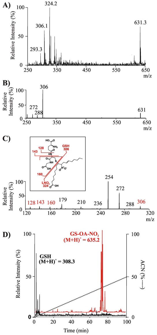
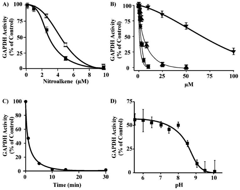
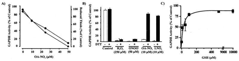
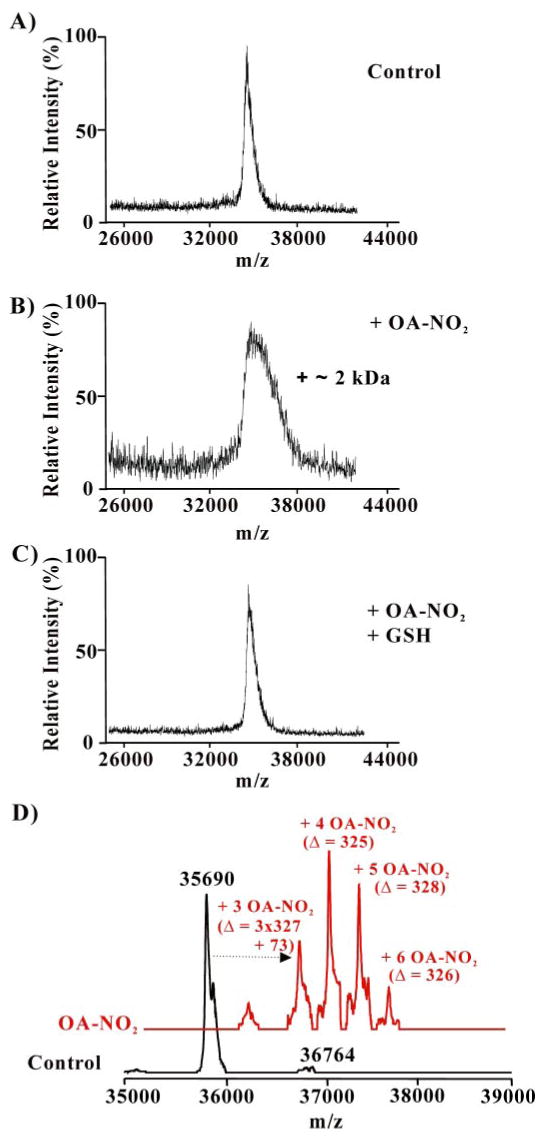
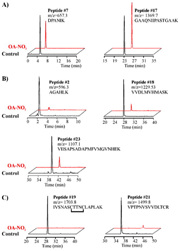
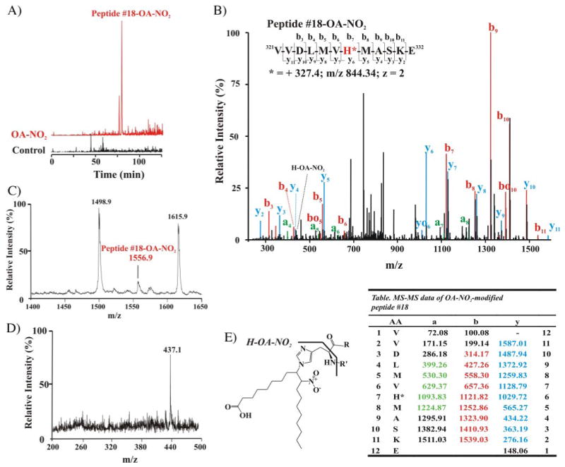
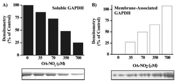
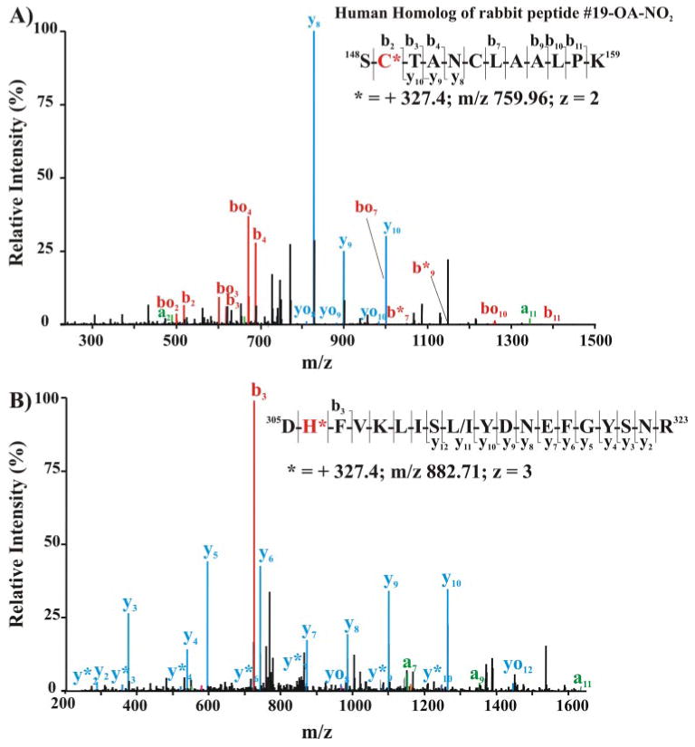
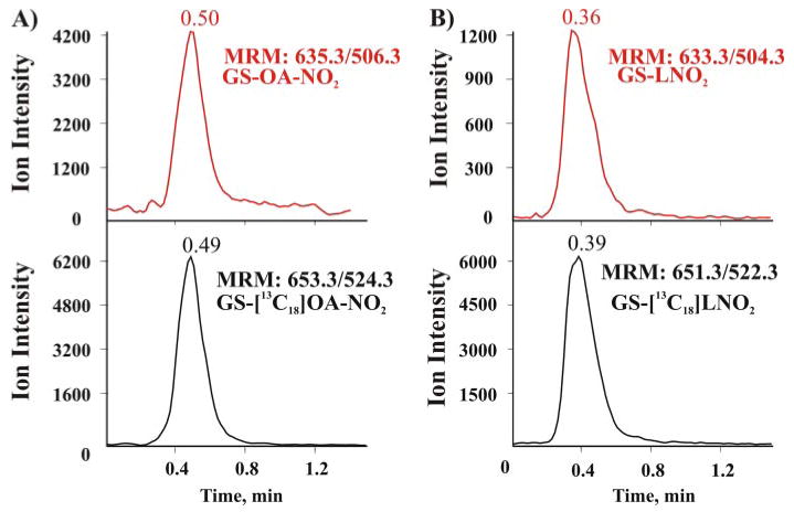
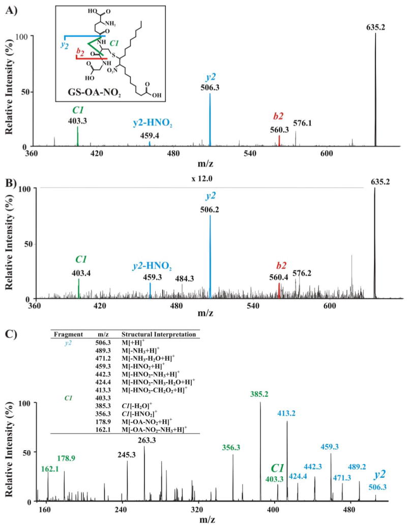
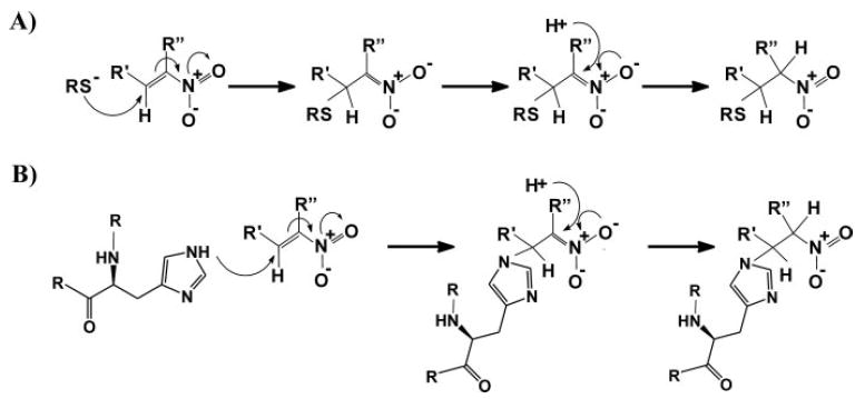
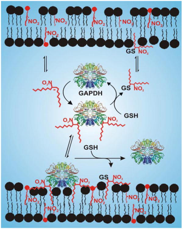
Nitric oxide ((*)NO)-derived reactive species nitrate unsaturated fatty acids, yielding nitroalkene derivatives, including the clinically abundant nitrated oleic and linoleic acids. The olefinic nitro group renders these derivatives electrophilic at the carbon beta to the nitro group, thus competent for Michael addition reactions with cysteine and histidine. By using chromatographic and mass spectrometric approaches, we characterized this reactivity by using in vitro reaction systems, and we demonstrated that nitroalkene-protein and GSH adducts are present in vivo under basal conditions in healthy human red cells. Nitro-linoleic acid (9-, 10-, 12-, and 13-nitro-9,12-octadecadienoic acids) (m/z 324.2) and nitro-oleic acid (9- and 10-nitro-9-octadecaenoic acids) (m/z 326.2) reacted with GSH (m/z 306.1), yielding adducts with m/z of 631.3 and 633.3, respectively. At physiological concentrations, nitroalkenes inhibited glyceraldehyde-3-phosphate dehydrogenase (GAPDH), which contains a critical catalytic Cys (Cys-149). GAPDH inhibition displayed an IC(50) of approximately 3 microM for both nitroalkenes, an IC(50) equivalent to the potent thiol oxidant peroxynitrite (ONOO(-)) and an IC(50) 30-fold less than H(2)O(2), indicating that nitroalkenes are potent thiol-reactive species. Liquid chromatography-mass spectrometry analysis revealed covalent adducts between fatty acid nitroalkene derivatives and GAPDH, including at the catalytic Cys-149. Liquid chromatography-mass spectrometry-based proteomic analysis of human red cells confirmed that nitroalkenes readily undergo covalent, thiol-reversible post-translational modification of nucleophilic amino acids in GSH and GAPDH in vivo. The adduction of GAPDH and GSH by nitroalkenes significantly increased the hydrophobicity of these molecules, both inducing translocation to membranes and suggesting why these abundant derivatives had not been detected previously via traditional high pressure liquid chromatography analysis. The occurrence of these electrophilic nitroalkylation reactions in vivo indicates that this reversible post-translational protein modification represents a new pathway for redox regulation of enzyme function, cell signaling, and protein trafficking.


Figures

FIGURE 1
FIGURE 1. Mass spectrometric analysis of the alkylation of glutathione by nitroalkenes and its impact on GSH lipophilicity
A, ESI-ion trap MS spectrum in negative ion mode (LCQ Deca; Thermo Electron Corp.) of the reaction product generated by LNO2 (300 μm) reaction with GSH (300 μm) in 50 mm sodium phosphate buffer, pH 7.4, at 20 °C for 30 min. Previous to the MS analysis, the reaction mixture was diluted in methanol, 0.1% NH4OH. B, MS/MS spectrum in negative ion mode of the GS-LNO2 adduct (m/z = 631.3). C, MS/MS/MS spectrum of fragment ion 306 m/z from GS-LNO2 adduct (m/z = 631.3). Inset, structural scheme of the adduct showing main fragmentation sites. The mass of main fragment ions is shown in red as detected in the negative ionization mode. D, total ion chromatogram in positive ion mode shows a significant increase in the lipophilicity of GSH following alkylation by OA-NO2, as indicated by the change in retention times of GSH ((M + H)+ = 308.3) (D, black) versus the GS-OA-NO2 adduct ((M + H)+ = 635.3) (D, red).
FIGURE 2
FIGURE 2. The inhibition of GAPDH following alkylation by nitrolinoleate, nitro-oleate, peroxynitrite, and hydrogen peroxide
A, GAPDH (0.5 μm) was incubated with increasing concentrations of LNO2 (0–10 μm) (☐) or OA-NO2 (■) in 100 mm sodium pyrophosphate, 100 μm DTPA, pH 7.4, at 20 °C for 15 min. Aliquots were removed, and GAPDH activity was determined. B, the relative inactivation of GAPDH by LNO2 (●), OA-NO2 (■), ONOO (0–50 μm; ▲), and H2O2 (0–100 μm, ▼). C, time course of OA-NO2-mediated GAPDH inhibition. GAPDH (0.5 μm) was incubated with OA-NO2 (10 μm) in 0.1 m pyrophosphate 0.1 mm DTPA, pH 7.4, at 25 °C. At the indicated time points aliquots were removed, and enzyme activity was determined. D, pH profile of the inhibition of GAPDH by OA-NO2. After preincubation of GAPDH (0.5 μm) for 5 min in 50 mm sodium pyrophosphate buffer adjusted to pH 5.5–10 at 20 °C, OA-NO2 (7.5 μm) was added, and after 15 min GAPDH activity was assessed as before. The percentage of control GAPDH activity at each pH was determined.
FIGURE 3
FIGURE 3. OA-NO2-induced thiol oxidation in GAPDH and reversibility of enzyme inactivation by thiol reagents
A, GAPDH (2 μm) was incubated for 15 min in 0.1 m pyrophosphate, 0.1 mm DTPA, pH 7.4, at 25 °C with OA-NO2 (0–50 μm). Aliquots were removed, and GAPDH activity was determined (♦). Reduced thiol content was determined by 5,5′-dithiobis(2-nitrobenzoic) acid reaction of GAPDH denatured in 1% SDS (●). B, GAPDH (0.5 μm) was incubated with H2O2 (250 μm), ONOO (50 μm), OA-NO2 (10 μm), or LNO2 (10 μm) in 0.1 m pyrophosphate, 0.1 mm DTPA, pH 7.4, at 25 °C for 15 min, and GAPDH activity was determined before (open bars) and after (solid black bars) treatment of the samples with DTT (10 mm) for 30 min at 25 °C. As a control, GAPDH was pretreated with DTT and activity was measured. C, OA-NO2-inactivated GAPDH was treated with increasing concentrations of GSH (0–10 mm) and enzyme activity determined.
FIGURE 4
FIGURE 4. Mass spectrometric analysis of GAPDH alkylation by OA-NO2 and its reversal by GSH
GAPDH (0.5 μm) was incubated with OA-NO2 (10 μm) in 0.1 m pyrophosphate, 0.1 mm DTPA, pH 7.4, at 25 °C for 15 min. GSH (10 mm) was then added for 15 min. After desalting, aliquots were analyzed by MALDI-TOF MS (AC; Voyager DE PRO, Applied Biosystems, Foster City, CA) or by LC-ESI two-dimensional linear ion trap MS (D; LTQ; Thermo Electron Corp.). A, spectrum of native GAPDH; B, OA-NO2-treated GAPDH before, and C, after addition of GSH; D, native GAPDH (black trace) and OA-NO2-treated GAPDH (red trace).
FIGURE 5
FIGURE 5. ESI-HPLC-MS analysis of tryptic peptides from native and OA-NO2-alkylated GAPDH
Selective ion chromatograms from native or OA-NO2-treated GAPDH digested with sequence-grade trypsin and analyzed by ESI-LC-MS (LCQ-Deca; Thermo Electron Corp.). Nonalkylated peptides (A) and peptides alkylated by OA-NO2 (B and C) are numbered as in the supplemental Fig. 1 and Table 1. A, similar relative ion intensities of non-nucleophilic peptides 7 and 17 (m/z 657.3 and 1369.7) were generated by both control and OA-NO2-nitroalkylated GAPDH. B, peptides 2, 18, and 23 containing the nucleophilic OA-NO2-reactive amino acid His were present at lower ion intensities in the tryptic digest of OA-NO2-treated GAPDH. C, peptides 19 and 21, containing OA-NO2-reactive nucleophilic amino acid Cys, were absent in tryptic digests of OA-NO2-treated GAPDH.
FIGURE 6
FIGURE 6. Mass spectrometric analysis of nitroalkylation patterns following in vitro treatment of purified GAPDH with OA-NO2
A, selective ion chromatograms from LC-ESI-MS analysis of OA-NO2-modified peptide 18 (LCQ Deca; Thermo Electron Corp.). The nitroalkylation of peptide 18 (native peptide (M + H)+ = 1229.6, room temperature 27.9 min) by OA-NO2 increased the retention time to 76 min, and the mass of the OA-NO2-modified peptide 18 was increased by 327 Da, equivalent to the neutral ion mass of OA-NO2, becoming m/z 1556.7. B, LC nanospray MS/MS spectrum of the nitroalkylated peptide 18 (LTQ; Thermo Electron Corp.). MS/MS spectrum of the doubly charged ion at m/z 844.34. Colors are annotated in the corresponding table. The yo and bo nomenclature indicates the corresponding y-H2O and b-H2O fragments, respectively. Inset, amino acid sequence of peptide 18 indicating major C- and N-terminal fragment ions detected by full-scan MS/MS. C, MALDI-TOF mass spectrum of the tryptic digest of OA-NO2-treated GAPDH (Voyager DE Pro, Applied Biosystem, Foster City, CA), focusing on nitroalkylated-peptide 18 ((M + H)+ = 1556.9). D, PSD MALDI-TOF-MS analysis of modified peptide 18 gives a main product ion at m/z 437.1, corresponding to the immonium ion of the histidine (H)-OA-NO2 adduct. E, structure and fragmentation pattern of the His-OA-NO2 adduct, showing the immonium adduct fragment (H-OA-NO2). Table list of MS/MS fragment ions m/z from peptide 18. Ions that are detected are highlighted in color (B). AA, amino acids.
FIGURE 7
FIGURE 7. Increased membrane association of GAPDH following nitroalkylation by OA-NO2
Control and OA-NO2-treated GAPDH were incubated with liposomes for 30 min at 25 °C. Liposomes were sedimented by ultracentrifugation, and the translocation of soluble GAPDH in the supernatant (A) to a liposome membrane-associated state (B) was determined as a function of OA-NO2 treatment concentration by SDS-PAGE.
FIGURE 8
FIGURE 8. Mass spectrometric detection of endogenous nitroalkylated GAPDH in red blood cells obtained from healthy humans
The cytosolic (A) and membrane-associated (B) protein fractions from lysed red cells were separated by SDS-PAGE using nonreducing, denaturing conditions (4–15% gradient gel). The 36-kDa Coomassie dye-binding band corresponding to the Rf of GAPDH was excised and digested in-gel with sequencing grade trypsin. Peptides were extracted, separated, and analyzed by LC nanospray linear ion trap MS/MS (LTQ; Thermo Electron Corp.). A, MS/MS of the doubly charged ion at m/z 759.96 corresponding to the human homolog of rabbit nitroalkylated peptide 19. Inset, amino acid sequence indicating major C- and N-terminal fragment ions detected by full-scan MS/MS. B, MS/MS spectrum of triply charged human GAPDH ion at m/z 882.71 corresponding to the human peptide sequence 305–323. Inset, amino acid sequence indicating major C- and N-terminal fragment ions detected by full-scan MS/MS.
FIGURE 9
FIGURE 9. Mass spectrometric detection of endogenous nitroalkylated GSH in healthy human red blood cells
Red cells were lysed, and membranes sedimented by centrifugation and the soluble fraction were supplemented with the internal standards GS-[13C18] OA-NO2 and GS-[13C18 (LNO2) before purification by reverse phase chromatography using a preparative C18 column. The eluted fraction was concentrated and analyzed by LC-ESI-MS/MS (Q-Trap 4000; Applied Biosystem, Foster City, CA). A, mass spectra of the eluent produced by monitoring the MRM transition 635.3/506.3 (corresponding to the generation of the y2-adducted fragment) for endogenous GS-OA-NO2 (red trace) and 653.3/524.3 for the added internal standard GS-[13C18] OA-NO2 (MRM 653.3/524.3 (black trace). B, similar to A, but monitoring the transitions for GS-LNO2. The injection peak in Fig. 9 occurred at 0.13 min.
FIGURE 10
FIGURE 10. Mass spectrometric characterization of nitroalkylated GSH
A, EPI analysis (e.g. MS/MS fragmentation pattern) of the synthetic standard GS-OA-NO2 showing major C- and N-terminal fragment ions detected by full-scan MS/MS (y and b fragments, respectively). Inset, scheme showing the structure and principal EPI fragments of GS-OA-NO2. B, EPI analysis of the endogenous RBC cytosolic GS-OA-NO2 adduct, displaying a fragmentation pattern identical to that of synthetic GS-OA-NO2. C, MS/MS/MS of the fragment ion y2 (m/z 506.3) from GS-OA-NO2 adduct (m/z = 635.2). Table, list and structural interpretation of fragment ions generated.
SCHEME 1
SCHEME 1. Michael addition reaction of fatty acid nitroalkene derivatives with (A) thiols and (B) amino groups
SCHEME 2
SCHEME 2. Nitroalkene-mediated post-translational modification of GAPDH and other proteins will influence protein structure, function, and subcellular distribution in a GSH-reversible manner
The modified sites of the protein were randomly assigned.

References

    1. Rubbo H, Darley-Usmar V, Freeman BA. Chem Res Toxicol. 1996;9:809–820. - PubMed
    1. Schopfer FJ, Baker PR, Giles G, Chumley P, Batthyany C, Crawford J, Patel RP, Hogg N, Branchaud BP, Lancaster JR, Jr, Freeman BA. J Biol Chem. 2005;280:19289–19297. - PubMed
    1. Napolitano A, Camera E, Picardo M, d'Ischia M. J Org Chem. 2000;65:4853–4860. - PubMed
    1. O'Donnell VB. Antioxid Redox Signal. 2003;5:195–203. - PubMed
    1. Baker PR, Lin Y, Schopfer FJ, Woodcock SR, Groeger AL, Batthyany C, Sweeney S, Long MH, Iles KE, Baker LM, Branchaud BP, Chen YE, Freeman BA. J Biol Chem. 2005;280:42464–42475. - PMC - PubMed

Publication types

MeSH terms