

A novel enzyme-linked immunosorbent assay for diagnosis of Mycobacterium avium subsp. paratuberculosis infections (Johne's Disease) in cattle
- PMID: 16682472
- PMCID: PMC1459654
- DOI: 10.1128/CVI.13.5.535-540.2006
A novel enzyme-linked immunosorbent assay for diagnosis of Mycobacterium avium subsp. paratuberculosis infections (Johne's Disease) in cattle
Abstract
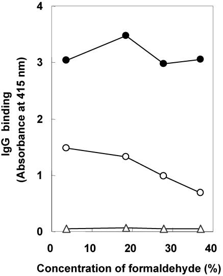
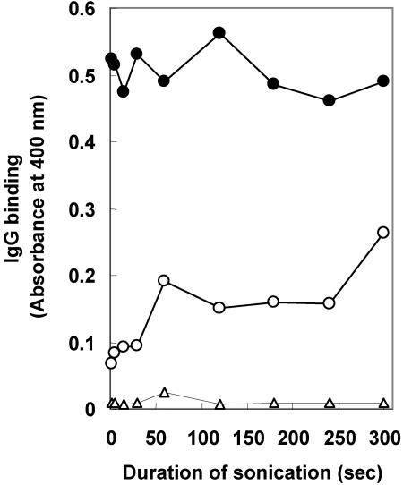
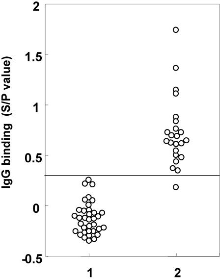
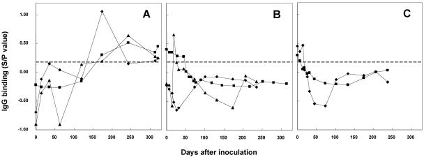
Enzyme-linked immunosorbent assays (ELISAs) for the diagnosis of Johne's disease (JD), caused by Mycobacterium avium subsp. paratuberculosis, were developed using whole bacilli treated with formaldehyde (called WELISA) or surface antigens obtained by treatment of M. avium subsp. paratuberculosis bacilli with formaldehyde and then brief sonication (called SELISA). ELISA plates were coated with either whole bacilli or sonicated antigens and tested for reactivity against serum obtained from JD-positive and JD-negative cattle or from calves experimentally inoculated with M. avium subsp. paratuberculosis, Mycobacterium avium subsp. avium, or Mycobacterium bovis. Because the initial results obtained from the WELISA and SELISA were similar, most of the subsequent experiments reported herein were performed using the SELISA method. To optimize the SELISA test, various concentrations (3.7 to 37%) of formaldehyde and intervals of sonication (2 to 300 s) were tested. With an increase in formaldehyde concentration and a decreased interval of sonication, there was a concomitant decrease in nonspecific binding by the SELISA. SELISAs prepared by treating M. avium subsp. paratuberculosis with 37% formaldehyde and then a 2-s burst of sonication produced the greatest difference (7x) between M. avium subsp. paratuberculosis-negative and M. avium subsp. paratuberculosis-positive serum samples. The diagnostic sensitivity and specificity for JD by the SELISA were greater than 95%. The SELISA showed subspecies-specific detection of M. avium subsp. paratuberculosis infections in calves experimentally inoculated with M. avium subsp. paratuberculosis or other mycobacteria. Based on diagnostic sensitivity and specificity, the SELISA appears superior to the commercial ELISAs routinely used for the diagnosis of JD.
Figures

References
-
- Brown, W. J., and M. V. G. Farquhar. 1989. Immunoperoxidase methods for the localization of antigens in cultured cells and tissue sections by electron microscopy. Methods Cell Biol. 31:553-569. - PubMed
-
- Chi, J., J. A. VanLeeuwen, A. Weersink, and G. P. Keefe. 2002. Direct production losses and treatment costs from bovine viral diarrhoea virus, bovine leukosis virus, Mycobacterium avium subsp. paratuberculosis and Neospora caninum. Prev. Vet. Med. 55:137-153. - PubMed
-
- Dubois, M., K. A. Gilles, J. K. Hamilton, P. A. Rebers, and F. Smith. 1956. Colorimetric methods for determination of sugars and related substances. Anal. Chem. 28:350-356.
-
- Eda, S., B. Elliott, M. C. Scott, W. R. Waters, J. P. Bannantine, R. H. Whitlock, and C. A. Speer. 2005. New method of serological testing for Mycobacterium avium subsp. paratuberculosis (Johne's disease) by flow cytometry. Foodborne Pathog. Dis. 2:250-262. - PubMed
-
- Koets, A. P., V. P. M. G. Rutten, M. de Boer, D. Bakker, P. Valentin-Weigand, and W. van Eden. 2001. Differential changes in heat shock protein-, lipoarabinomannan-, and purified protein derivative-specific immunoglobulin G1 and G2 isotype responses during bovine Mycobacterium avium subsp. paratuberculosis infection. Infect. Immun. 69:1492-1498. - PMC - PubMed
Publication types
MeSH terms
Substances
LinkOut - more resources
Full Text Sources
