. 2006 May 15;203(5):1273-82.
doi: 10.1084/jem.20052205. Epub 2006 May 8.

Lymphocyte recruitment into the aortic wall before and during development of atherosclerosis is partially L-selectin dependent

Affiliations

Lymphocyte recruitment into the aortic wall before and during development of atherosclerosis is partially L-selectin dependent

Elena Galkina et al. J Exp Med. .

Abstract

Atherosclerosis is an inflammatory disease of large arteries. Flow cytometry of aortic cell suspensions showed that B and T lymphocytes and some macrophages and dendritic cells are already present in the adventitia of normal/noninflamed mouse aortas. Adoptively transferred lymphocytes constitutively homed to the aorta and resided within the adventitia up to 7 d after transfer. Lymphocyte trafficking into normal/noninflamed or atherosclerosis-prone aortas was partially L-selectin dependent. Antigen-activated dendritic cells induced increased T lymphocyte proliferation within the aorta 72 h after adoptive transfer. During progression of atherosclerosis in apolipoprotein-E-deficient mice, the total number of macrophages, T cells, and dendritic cells, but not B cells, increased significantly. This alteration in immune cell composition was accompanied by the formation of tertiary lymphoid tissue in the adventitia of atherosclerotic aortas. These results demonstrate that lymphocytes already reside within the normal/noninflamed aorta before the onset atherosclerosis as a consequence of constitutive trafficking. Atherosclerosis induces the recruitment of macrophages and dendritic cells that support antigen presentation.


Figures
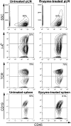
Figure 1.
Figure 1.
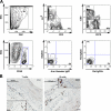
Method validation for flow cytometry analysis of mouse aorta. Cell suspensions from pLN or spleen, untreated or treated with enzyme cocktail were stained with anti-CD45 (PE-Texas red), anti–I-Ab (PE), anti-TCRβ (APC), and anti-CD19 (PE-Cy7) Abs; numbers in the gate represent the percentage of CD45+ cells of all analyzed cells. The numbers in the top right quadrant of the bottom six panels represent the percentages of I-Ab+ , TCRβ+, CD19+ cells among all CD45+ cells.
Figure 2.
Figure 2.
T and B cells reside within normal/noninflamed aortic wall of C57BL/6 mice. (A) Leukocytes within the aortic wall of C57BL/6 mice. Aortas from C57BL/6 mice were perfused by cardiac puncture with phosphate-buffered heparin. Collected aortas were digested in enzymes for 1 h at 37°C and cell suspensions were stained with anti-CD45, -CD3, -CD19, -CD5, and I-Ab Abs or with isotype control Abs. To eliminate autofluorescence from debris and necrotic tissues, R1 gate (FSC < 30,000, FSC > 500) was applied for all samples. Then CD45+ cells (gate R2) were defined within R1 gate and further analysis was performed on R1 and R2 cells. (B) B and T cells reside within the aortic wall of C57BL/6 mice. C57BL/6 mice were perfused by cardiac puncture with phosphate-buffered heparin and fixed with 4% paraformaldehyde (PFA). Paraffin-embedded longitudinal sections were stained with anti-CD3 (T cells) or anti-CD20 (B cells) Abs with avidin-biotin technology and counterstained with hematoxylin. Isotype-control Abs showed no staining (not depicted).
Figure 3.
Figure 3.
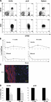
Constitutive lymphocyte homing into the normal/noninflamed aortic wall. (A) Adoptive transfer of splenocytes from C57BL/6 mice into Rag-1 −/− recipient mice. CFSE-labeled splenocytes from C57BL/6 mice were injected i.v. into Rag-1 −/− mice. After 24 h, cell suspensions from aorta, spleen, and pLN were obtained and stained with Abs against CD3, CD19, and CD45. The top row is gated on CD45+; the bottom row is gated on CFSE+CD19. Numbers in quadrants are percentage of positive cells. (B) Kinetics of CFSE-labeled lymphocyte migration into Rag-1 −/− mice (n = 3–9 mice, mean ± SE). C57BL/6 lymphocytes were labeled with CFSE and 40 × 106 lymphocytes were injected i.v. into Rag-1 −/− recipient mice. Aorta, blood, spleen, and pLN were collected at 20 min and 1, 3, and 7 d after adoptive transfer; cell suspensions from different organs were stained with anti-CD45, CD19, TCR; and the percentages of CFSE-positive emigrated T and B lymphocytes were determined by flow cytometry. (C) Localization of CFSE-labeled lymphocytes 24 h after adoptive transfer (green-CFSE, blue-DAPI staining). (D) L-selectin–dependent lymphocyte homing into the aorta. C57BL/6 and L-selectin −/− splenocytes were labeled with either CFSE or CMTMR, mixed in equal numbers, and injected i.v. into recipient mice. Aortas (left) and LN (right) were collected after 24 h and stained with Abs against CD3, CD19, and CD45+. Homing of L-selectin −/− lymphocytes (gray bars) is expressed as a percent of C57BL/6 lymphocyte homing (100%, black bars). Results show mean ± SE from at least four recipients from at least three independent experiments. **, P < 0.01; ***, P < 0.001.
Figure 4.
Figure 4.
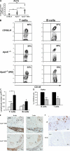
T and B lymphocyte compartment alterations during atherosclerosis. (A) Total leukocyte number in the aorta of 33–35 wk old C57BL/6, ApoE −/− and ApoE −/− (WD for 20 wk) mice. Cells were liberated from the aortic tissues and the total leukocyte number was determined (n = 14–23 mice). Mean is indicated by horizontal bar. *, P < 0.05; **, P < 0.01. (B) T and B lymphocyte compartments in C57BL/6, ApoE −/− and ApoE −/− (WD) mice. Cell suspensions from aortas of C57BL/6 (n = 12, white bar), ApoE −/− (n = 26, gray bar) and ApoE −/− (WD) (n = 21, black bar) mice were stained with Abs against CD45, TCRβ (left), and CD19 (right). The number in the quadrants represents the percentage of positive cells. All plots were gated on CD45+ cells. (C) Total T and B lymphocyte numbers in the aorta of C57BL/6, ApoE −/− and ApoE −/− (WD) mice were calculated from the total cell number and the percentage of each population. Results are means ± SE (n = 12–26). **, P < 0.01. (D) L-selectin–dependent lymphocyte homing into the atherosclerotic aorta. C57BL/6 and L-selectin −/− splenocytes were labeled with either CFSE or CMTMR, mixed in equal number, and injected i.v. into ApoE −/− (WD) recipient mice. Aortas were collected after 24 h and stained with Abs against CD3, CD19, and CD45. Homing of L-selectin −/− lymphocytes (gray bars) is expressed as the percentage of C57BL/6 lymphocyte homing (100%, black bars). Results show mean ± SE from at least four recipients from at least three independent experiments. **, P < 0.01. (E) B (left) and T (right) cells form organized lymphoid tissue in the adventitia of ApoE −/− (WD) mice. Paraffin-embedded sections (5 μm) were stained with Abs against CD20 and CD3, respectively, and counterstained with hematoxylin. Isotype-control Abs showed no staining (not depicted). L, neointimal lesion. (F) MECA-79 is expressed in LN (top), but not in lymphoid tissue in the aortas of ApoE −/− (WD) mice. Isotype-control Abs showed no staining (not depicted).
Figure 5.
Figure 5.
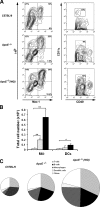
Immune cell composition during the development and progression of atherosclerosis. (A) Macrophages and dendritic cells in C57BL/6, ApoE −/− and ApoE −/− (WD) mice. Cell suspensions from mouse aortas were stained with Abs against CD45, I-Ab, Mac-1 (left) and CD11c and CD40 (right). Plots gated on CD45+ cells (macrophages) and on CD45+/CD40+ (DCs). Representative plots from one experiment are shown. (B) Total number of macrophages and dendritic cells in the aortas of C57BL/6 (white bars), ApoE −/− (gray bars), and ApoE −/− (WD) (black bars) mice, calculated based on the percentage of each population and the total number of leukocytes from the aortas. Results mean ± SE (n = 7–17). *, P < 0.05; **, P < 0.01. (C) Alterations in the immune cell composition during development and progression of atherosclerosis. The area of each circle is proportional to the number of CD45+ cells.
Figure 6.
Figure 6.
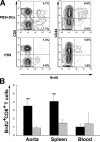
Immunization with antigen-activated DCs leads to proliferation of adventitial T lymphocytes within the aorta. CD40L-activated DCs were pulsed with antigen peptide and either 105 bone marrow–derived DCs in 200 μl PBS or 200 μl PBS were injected i.v. into OT-1 transgenic recipient mice. At 48 and 60 h, 1 mg BrdU was injected i.p. Aorta, pLN, spleen, and blood were collected 72 h after transfer, and the cell suspensions were stained with anti-CD45, -CD8, -CD44, and anti-BrdU Abs. All plots were gated on CD45+ lymphocyte gate. (A) Representative plots from one experiment. The numbers in quadrants represents BrdU+ cells. (B) The percentage of BrdU+ CD8+ T cells in the aortas and spleens of OT-1 control (gray bars) or immunized OT-1 recipient mice (black bars) are presented. Results mean ± SE (n = 8). **, P < 0.01.

Similar articles

Cited by

References

    1. Libby, P. 2002. Inflammation in atherosclerosis. Nature. 420:868–874. - PubMed
    1. Ross, R. 1999. Atherosclerosis–an inflammatory disease. N. Engl. J. Med. 340:115–126. - PubMed
    1. Hansson, G.K. 2005. Inflammation, atherosclerosis, and coronary artery disease. N. Engl. J. Med. 352:1685–1695. - PubMed
    1. Jonasson, L., J. Holm, O. Skalli, G. Bondjers, and G.K. Hansson. 1986. Regional accumulations of T cells, macrophages, and smooth muscle cells in the human atherosclerotic plaque. Arteriosclerosis. 6:131–138. - PubMed
    1. Buono, C., H. Pang, Y. Uchida, P. Libby, A.H. Sharpe, and A.H. Lichtman. 2004. B7-1/B7-2 costimulation regulates plaque antigen-specific T-cell responses and atherogenesis in low-density lipoprotein receptor-deficient mice. Circulation. 109:2009–2015. - PubMed

Publication types

MeSH terms