. 2006 May 16;103(20):7655-9.
doi: 10.1073/pnas.0601037103. Epub 2006 May 8.

Pannexin 1 in erythrocytes: function without a gap

Affiliations

Pannexin 1 in erythrocytes: function without a gap

Silviu Locovei et al. Proc Natl Acad Sci U S A. .

Abstract

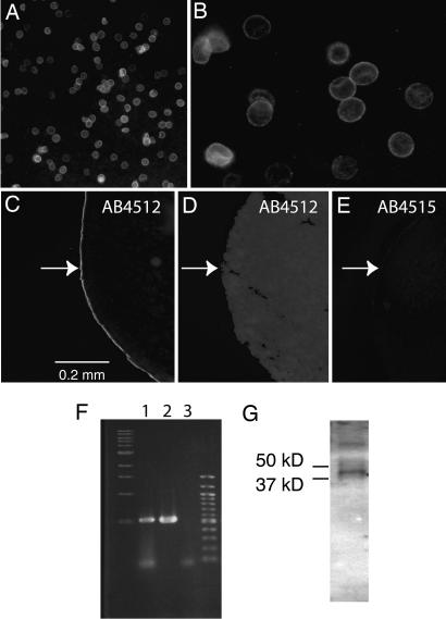
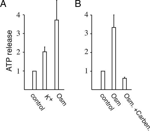
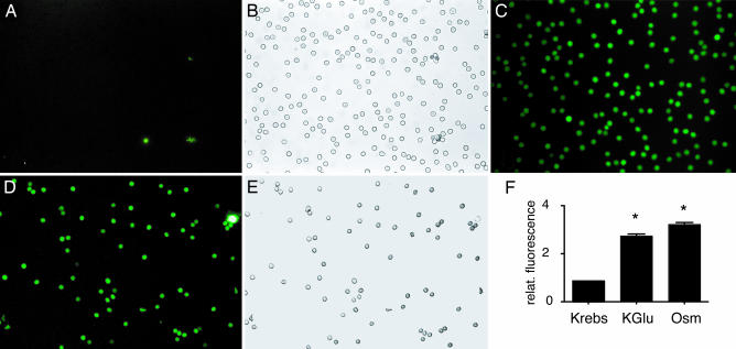
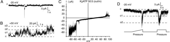
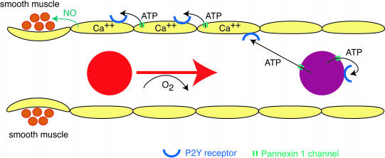
ATP is a widely used extracellular signaling molecule. The mechanism of ATP release from cells is presently unresolved and may be either vesicular or channel-mediated. Erythrocytes release ATP in response to low oxygen or to shear stress. In the absence of vesicles, the release has to be through channels. Erythrocytes do not form gap junctions. Yet, here we show with immunohistochemical and electrophysiological data that erythrocytes express the gap junction protein pannexin 1. This protein, in addition to forming gap junction channels in paired oocytes, can also form a mechanosensitive and ATP-permeable channel in the nonjunctional plasma membrane. Consistent with a role of pannexin 1 as an ATP release channel, ATP release by erythrocytes was attenuated by the gap junction blocker carbenoxolone. Furthermore, under conditions of ATP release, erythrocytes took up fluorescent tracer molecules permeant to gap junction channels.


Conflict of interest statement

Conflict of interest statement: No conflicts declared.

Figures

Fig. 1.
Fig. 1.
Expression of pannexin 1. (A) Human erythrocytes are stained by extracellular application of antibody 4512. (B) Antibody-reacted erythrocytes at higher magnification. (C and E) Extracellular application of antibody 4512 (C) but not 4515 (E) to intact oocytes expressing human pannexin 1 yielded immunostaining of the oocyte surface. Application of antibody 4515 to sectioned oocytes resulted in the same staining pattern as in C (Fig. 6). (D) Uninjected oocytes did not yield staining with either antibody, irrespective of the side of application. The arrows indicate the location of the oocyte surface. (F) PCR amplification of a human bone marrow cDNA library with pannexin 1-specific primers yielded the same size amplicon (lane 1) as seen with the pannexin 1 clone used for exogenous expression (lane 2); no amplicon was generated without addition of DNA (lane 3) to the PCR. Lanes 1–3 are flanked by size markers. (G) Western blot of human erythrocyte membranes with antibody 4512 yielded a band of expected size for pannexin 1. The higher-molecular-weight band may represent a dimer of the protein. Size markers were run on parallel lanes.
Fig. 2.
Fig. 2.
ATP release from human erythrocytes. ATP was measured with a luciferase assay in the supernatant after centrifugation. Data are normalized to basal release in unstimulated erythrocytes. (A) Release was increased by depolarization with potassium as well as by hypotonic stress with 1:1-diluted Krebs solution. (B) Inhibition of osmotically induced ATP release by 100 μM carbenoxolone. Means ± SEM are plotted (n = 5).
Fig. 3.
Fig. 3.
Uptake of the fluorescent tracer molecule carboxyfluorescein by human erythrocytes under conditions of ATP release. (A and B) In Krebs solution, fluorescence was only associated with cell debris or erythrocyte ghosts. (C) All erythrocytes in the field of view were fluorescent after stimulation of ATP release with high K+. (D and E) Low oxygen exposure yielded dye uptake in all erythrocytes in the field of view. (F) Quantitative analysis of tracer uptake induced by depolarization (KGlu) or osmotically (Osm) by incubating erythrocytes in 1:1-diluted Krebs solution. Means ± SEM are plotted (n = 5); P < 0.01 versus control (Krebs).
Fig. 4.
Fig. 4.
Channel currents in membrane patches excised from human erythrocytes with properties consistent with those of pannexin 1 channels. (A and B) Channel conductances of the magnitude of pannexin 1 channels were rarely seen at negative holding potentials (A) but were frequent at positive holding potentials (B). (C) The reversal potentials for membrane currents in a potassium ATP gradient (50 mM out/5 mM in) were of the same magnitude as those observed in pannexin 1 channels expressed in oocytes (3). (D) Like pannexin 1 channels, the large conductance channels in erythrocytes were mechanosensitive. The lines in B and D indicate current levels expected for full openings of pannexin 1 channels.
Fig. 5.
Fig. 5.
Scheme depicting a possible involvement of pannexin 1 channels in local blood-flow regulation. Oxygen-deprived or shear-stressed erythrocytes release ATP, which binds to P2Y receptors on erythrocytes and on endothelial cells. In erythrocytes, activation of the receptor leads to further release of ATP through pannexin 1 channels (ATP-induced ATP release). In endothelial cells, binding of ATP to P2Y receptors or shear stress initiates a calcium wave by opening pannexin 1 channels. The wave propagates retrogradely and reaches the precapillary sphincter region. Endothelial NO synthase is activated, and NO released from the endothelial cell relaxes the smooth muscle. In arterioles, the smooth muscle would be covering all endothelial cells and could be relaxed by NO without the need of a propagated wave.

References

    1. Panchin Y., Kelmanson I., Matz M., Lukyanov K., Usman N., Lukyanov S. Curr. Biol. 2000;10:R473–R474. - PubMed
    1. Bruzzone R., Hormuzdi S. G., Barbe M. T., Herb A., Monyer H. Proc. Natl. Acad. Sci. USA. 2003;100:13644–13649. - PMC - PubMed
    1. Bao L., Locovei S., Dahl G. FEBS Lett. 2004;572:65–68. - PubMed
    1. Burnstock G., Knight G. E. Int. Rev. Cytol. 2004;240:31–304. - PubMed
    1. Bal-Price A., Moneer Z., Brown G. C. Glia. 2002;40:312–323. - PubMed

Publication types