

Crystallogenesis of bacteriophage P22 tail accessory factor gp26 at acidic and neutral pH
- PMID: 16682781
- PMCID: PMC2219973
- DOI: 10.1107/S1744309106013856
Crystallogenesis of bacteriophage P22 tail accessory factor gp26 at acidic and neutral pH
Abstract
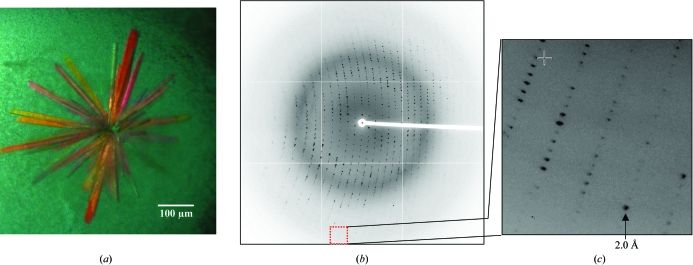
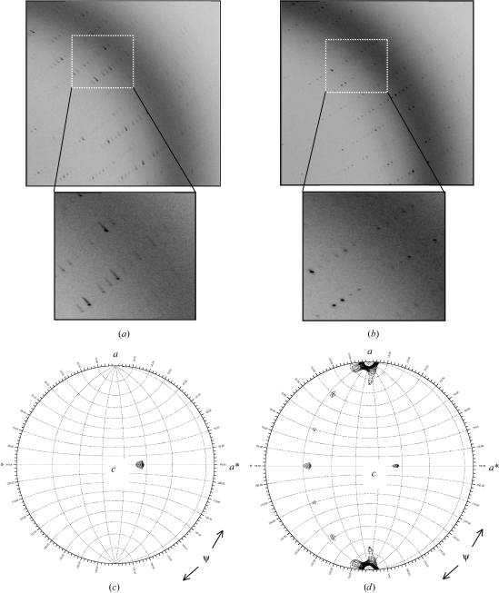
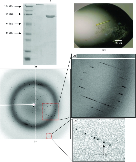
Gp26 is one of three phage P22-encoded tail accessory factors essential for stabilization of viral DNA within the mature capsid. In solution, gp26 exists as an extended triple-stranded coiled-coil protein which shares profound structural similarities with class I viral membrane-fusion protein. In the cryo-EM reconstruction of P22 tail extracted from mature virions, gp26 forms an approximately 220 angstroms extended needle structure emanating from the neck of the tail, which is likely to be brought into contact with the cell's outer membrane when the viral DNA-injection process is initiated. To shed light on the potential role of gp26 in cell-wall penetration and DNA injection, gp26 has been crystallized at acidic, neutral and alkaline pH. Crystals of native gp26 grown at pH 4.6 diffract X-rays to 2.0 angstroms resolution and belong to space group P2(1), with a dimer of trimeric gp26 molecules in the asymmetric unit. To study potential pH-induced conformational changes in the gp26 structure, a chimera of gp26 fused to maltose-binding protein (MBP-gp26) was generated. Hexagonal crystals of MBP-gp26 were obtained at neutral and alkaline pH using the high-throughput crystallization robot at the Hauptman-Woodward Medical Research Institute, Buffalo, NY, USA. These crystals diffract X-rays to beyond 2.0 angstroms resolution. Structural analysis of gp26 crystallized at acidic, neutral and alkaline pH is in progress.
Figures
Similar articles
-
Structural Plasticity of the Protein Plug That Traps Newly Packaged Genomes in Podoviridae Virions.J Biol Chem. 2016 Jan 1;291(1):215-26. doi: 10.1074/jbc.M115.696260. Epub 2015 Nov 16. J Biol Chem. 2016. PMID: 26574546 Free PMC article.
-
Bacteriophage P22 tail accessory factor GP26 is a long triple-stranded coiled-coil.J Biol Chem. 2005 Feb 18;280(7):5929-33. doi: 10.1074/jbc.C400513200. Epub 2004 Dec 8. J Biol Chem. 2005. PMID: 15591072
-
Structure of phage P22 cell envelope–penetrating needle.Nat Struct Mol Biol. 2007 Dec;14(12):1221-6. doi: 10.1038/nsmb1317. Nat Struct Mol Biol. 2007. PMID: 18059287
-
Phage tailspike protein. A fishy tale of protein folding.Curr Biol. 1994 Nov 1;4(11):1026-9. doi: 10.1016/s0960-9822(00)00234-7. Curr Biol. 1994. PMID: 7874487 Review.
-
There's a right way and a wrong way: in vivo and in vitro folding, misfolding and subunit assembly of the P22 tailspike.Structure. 1999 Jun 15;7(6):R131-9. doi: 10.1016/s0969-2126(99)80078-1. Structure. 1999. PMID: 10404587 Review.
Cited by
-
Structural Plasticity of the Protein Plug That Traps Newly Packaged Genomes in Podoviridae Virions.J Biol Chem. 2016 Jan 1;291(1):215-26. doi: 10.1074/jbc.M115.696260. Epub 2015 Nov 16. J Biol Chem. 2016. PMID: 26574546 Free PMC article.
-
Exploring the atomic structure and conformational flexibility of a 320 Å long engineered viral fiber using X-ray crystallography.Acta Crystallogr D Biol Crystallogr. 2014 Feb;70(Pt 2):342-53. doi: 10.1107/S1399004713027685. Epub 2014 Jan 29. Acta Crystallogr D Biol Crystallogr. 2014. PMID: 24531468 Free PMC article.
-
Evolution of Lactococcus lactis phages within a cheese factory.Appl Environ Microbiol. 2009 Aug;75(16):5336-44. doi: 10.1128/AEM.00761-09. Epub 2009 Jun 19. Appl Environ Microbiol. 2009. PMID: 19542338 Free PMC article.
-
Structural plasticity of the phage P22 tail needle gp26 probed with xenon gas.Protein Sci. 2009 Mar;18(3):537-48. doi: 10.1002/pro.53. Protein Sci. 2009. PMID: 19241380 Free PMC article.
References
-
- Andrews, D., Butler, J. S., Al-Bassam, J., Joss, L., Winn-Stapley, D. A., Casjens, S. & Cingolani, G. (2005). J. Biol. Chem.280, 5929–5933. - PubMed
-
- Bazinet, C., Benbasat, J., King, J., Carazo, J. M. & Carrascosa, J. L. (1988). Biochemistry, 27, 1849–1856. - PubMed
-
- Casjens, S. (1979). J. Mol. Biol.131, 1–14. - PubMed
-
- Casjens, S. & Huang, W. M. (1982). J. Mol. Biol.157, 287–298. - PubMed
-
- Casjens, S. & King, J. (1974). J. Supramol. Struct.2, 202–224. - PubMed
MeSH terms
Substances
LinkOut - more resources
Full Text Sources
Miscellaneous
