. 2006 May 9:7:21.
doi: 10.1186/1471-2121-7-21.

Pim-1 kinase phosphorylates RUNX family transcription factors and enhances their activity

Affiliations

Pim-1 kinase phosphorylates RUNX family transcription factors and enhances their activity

Teija L T Aho et al. BMC Cell Biol. .

Abstract

Background: The pim family genes encode oncogenic serine/threonine kinases which in hematopoietic cells have been implicated in cytokine-dependent signaling as well as in lymphomagenesis, especially in cooperation with other oncogenes such as myc, bcl-2 or Runx family genes. The Runx genes encode alpha-subunits of heterodimeric transcription factors which regulate cell proliferation and differentiation in various tissues during development and which can become leukemogenic upon aberrant expression.

Results: Here we have identified novel protein-protein interactions between the Pim-1 kinase and the RUNX family transcription factors. Using the yeast two-hybrid system, we were able to show that the C-terminal part of human RUNX3 associates with Pim-1. This result was confirmed in cell culture, where full-length murine Runx1 and Runx3 both coprecipitated and colocalized with Pim-1. Furthermore, catalytically active Pim-1 kinase was able to phosphorylate Runx1 and Runx3 proteins and enhance the transactivation activity of Runx1 in a dose-dependent fashion.

Conclusion: Altogether, our results suggest that mammalian RUNX family transcription factors are novel binding partners and substrates for the Pim-1 kinase, which may be able to regulate their activities during normal hematopoiesis as well as in leukemogenesis.


Figures
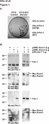
Figure 1
Figure 1
Pim-1 interacts with RUNX family proteins. (A) Yeast strains expressing the VP16 activation domain alone or fused with the B19 fragment of human RUNX3 were mated with strains expressing the GAL4 DNA-binding domain fused with the control protein lamin or either kinase-deficient (K67M) or wild-type (WT) Pim-1. The ability of two proteins to interact with each other was judged based on the capacity ot the corresponding diploid strains to grow on the selective medium lacking histidine. (B) COS-7 cells were transfected with pSV-pim-1, pAMC-Runx1 or pAMC-Runx3 plasmids as indicated in the figure. Parts of the cell lysates were subjected to immunoprecipitation with anti-Myc antibody followed by immunoblotting with anti-Pim-1 or anti-Myc antibodies. The expression of proteins in the lysates was verified by direct Western blotting with the same antibodies.
Figure 2
Figure 2
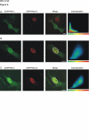
Pim-1 colocalizes with Runx-1 and Runx-3. The subcellular distribution of ECFP-Pim-1, EYFP-Runx1 and EYFP-Runx3 was analysed from transiently transfected COS-7 cells under confocal microscope. Shown are single- (A) or double-positive (B, C) cells expressing indicated fluorescent proteins. Colocalization of ECFP (first panel) and EYFP (second panel) fusion proteins in the circled nuclei was visualized by yellow colour in merged images (third panel) and was confirmed by scattergram plots (fourth panel), where the intensities of the CFP and YFP channels are on the X- and Y-axis, respectively. Bar represents 20 μm.
Figure 3
Figure 3
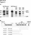
Pim-1 phosphorylates Runx proteins in vitro. (A) Bacterially produced GST fusion proteins expressing either full-length (FL) or fragments of Runx1 or Runx3 were incubated with GST-Pim-1 in in vitro kinase assays. The phosphorylation products were separated on SDS-PAGE and visualized by autoradiography. GST alone (-) was used as a negative control. * indicates protein degradation products. (B) Schematic presentation of the functional domains of Runx1, including the Runt domain, an activation domain (AD) with two major transactivation elements (TE1 and TE2), a minor transactivation element (TE3), an inhibitory domain (ID) and the C-terminal VWRPY sequence. Shown are also the fragments phosphorylated by Pim-1 in Runx1 or Runx3, which lacks the sequences corresponding to TE3 of Runx1.
Figure 4
Figure 4
Pim-1 potentiates transcriptional activity of Runx1. (A) Jurkat TAg-cells were transfected with 4 μg of pM-CSF-R-Luc, 1 μg of pSV-β-gal, 4 μg of pEF-Runx1, 2 μg of pEF-Cbfβ2, and indicated amounts of pLTR-pim-1. The steady-state levels of Pim-1 protein were measured from the same cell lysates by Western blotting with anti-Pim-1 antibody and equal loading was verified with anti-β-actin antibody. (B) Jurkat TAg-cells were transfected with same reporter constructs as in Figure A together with wild-type or mutant pSV-pim-1 constructs. (C) Jurkat TAg-cells were transfected with 3 μg of pG5-Luc, 1 μg of pSV-β-gal, 3 μg of GAL4 fusion proteins and 2 μg of pEF-Cbfβ2 together with indicated amounts of pLTR-pim-1. Shown are relative luciferase activities normalized against β-galactosidase activities and statistically analysed by Student's t-test (*, p ≤ 0.05; **, p ≤ 0.01).

References

    1. Allen JD, Berns A. Complementation tagging of cooperating oncogenes in knockout mice. Semin Cancer Biol. 1996;7:299–306. doi: 10.1006/scbi.1996.0038. Review. - DOI - PubMed
    1. Acton D, Domen J, Jacobs H, Vlaar M, Korsmeyer S, Berns A. Collaboration of pim -1 and bcl -2 in lymphomagenesis. Curr Top Microbiol Immunol. 1992;182:293–298. - PubMed
    1. Blyth K, Terry A, Mackay N, Vaillant F, Bell M, Cameron ER, Neil JC, Stewart M. Runx2: a novel oncogenic effector revealed by in vivo complementation and retroviral tagging. Oncogene. 2001;20:295–302. doi: 10.1038/sj.onc.1204090. - DOI - PubMed
    1. Saris CJM, Domen J, Berns A. The pim-1 oncogene encodes two related protein-serine/threonine kinases by alternative initiation at AUG and CUG. EMBO J. 1991;10:655–664. - PMC - PubMed
    1. Dautry F, Weil D, Yu J, Dautry-Varsat A. Regulation of pim and myb mRNA accumulation by interleukin 2 and interleukin 3 in murine hematopoietic cell lines. J Biol Chem. 1988;263:17615–17620. - PubMed

Publication types

MeSH terms

Substances

LinkOut - more resources