Review
. 2006 May 9;7(1):73.
doi: 10.1186/1465-9921-7-73.

Muscarinic receptor signaling in the pathophysiology of asthma and COPD

Affiliations
Review

Muscarinic receptor signaling in the pathophysiology of asthma and COPD

Reinoud Gosens et al. Respir Res. .

Abstract

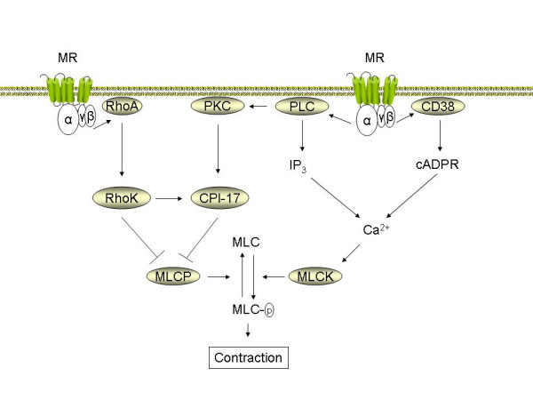
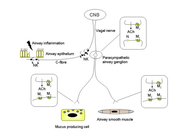
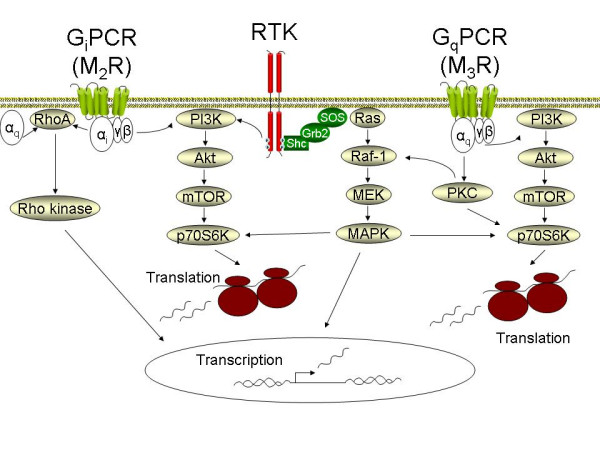
Anticholinergics are widely used for the treatment of COPD, and to a lesser extent for asthma. Primarily used as bronchodilators, they reverse the action of vagally derived acetylcholine on airway smooth muscle contraction. Recent novel studies suggest that the effects of anticholinergics likely extend far beyond inducing bronchodilation, as the novel anticholinergic drug tiotropium bromide can effectively inhibit accelerated decline of lung function in COPD patients. Vagal tone is increased in airway inflammation associated with asthma and COPD; this results from exaggerated acetylcholine release and enhanced expression of downstream signaling components in airway smooth muscle. Vagally derived acetylcholine also regulates mucus production in the airways. A number of recent research papers also indicate that acetylcholine, acting through muscarinic receptors, may in part regulate pathological changes associated with airway remodeling. Muscarinic receptor signalling regulates airway smooth muscle thickening and differentiation, both in vitro and in vivo. Furthermore, acetylcholine and its synthesizing enzyme, choline acetyl transferase (ChAT), are ubiquitously expressed throughout the airways. Most notably epithelial cells and inflammatory cells generate acetylcholine, and express functional muscarinic receptors. Interestingly, recent work indicates the expression and function of muscarinic receptors on neutrophils is increased in COPD. Considering the potential broad role for endogenous acetylcholine in airway biology, this review summarizes established and novel aspects of muscarinic receptor signaling in relation to the pathophysiology and treatment of asthma and COPD.


Figures

Figure 1
Figure 1
Pathways central in muscarinic receptor mediated airway smooth muscle contraction. Muscarinic receptor (MR) agonists induce contraction of airway smooth muscle by Ca2+ dependent and Ca2+ independent pathways. Through associated Gq alpha subunits, the muscarinic M3 receptor activates phospholipase C (PLC), which releases inositol 1,4,5-trisphosphate (IP3) and diacylglycerol (DAG) after hydrolytic conversion of phosphatidylinositol-4,5-bisphosphate (PIP2). IP3 induces the release of Ca2+ from internal sarcoplasmatic reticulum (SR) stores. Coupling of M3 receptor to CD38 through as yet undefined mechanisms contributes to the production of cyclic ADP ribose (cADPR) and the release of Ca2+ through ryanodine receptor channels in the SR. Ca2+ release increases free cytosolic Ca2+ and promotes calmodulin-dependent activation of myosin light chain kinase (MLCK). MLCK mediated phosphorylation of 20 kDa regulatory myosin light chain (MLC) in the contractile apparatus is an obligatory event to induce smooth muscle contraction. MLC phosphorylation level is also controlled by pathways that inhibit myosin light chain phosphatase (MLCP) and, thus enhance Ca2+ sensitivity. PLC-derived DAG activates protein kinase C (PKC), leading to CPI-17 phosphorylation and downstream MLCP inhibition. Rho-kinase, which is activated by the monomeric G protein RhoA, both phosphorylates CPI-17 and inhibits MLCP directly. The expression and function of RhoA, CPI-17 and CD38 are increased by pro-inflammatory cytokines in vitro and in animal models of asthma and COPD ex vivo (see text).
Figure 2
Figure 2
Cholinergic receptors involved in neuronal acetylcholine release and function. Neuronal acetylcholine release is regulated by a network of afferent and efferent airway nerves that interact with their surrounding cells. Afferent C-fibers project to the subepithelial region where they can be activated by inflammatory mediators and non-specific stimuli. In asthma, epithelial damage can expose sensory nerve endings to the airway lumen, potentiating their activation. Activated C-fibers secrete neurokinins (NK) that exert local effects and facilitate ganglionic neurotransmission (peripheral reflex arc). In addition, the activated C-fiber increases the output of the vagal nerve through regulation in the central nervous system (CNS) (central reflex arc). Neurotransmission in parasympathetic ganglia of the airway is mediated by acetylcholine through nicotinic (N) and muscarinic M1 receptors and can be markedly facilitated by inflammatory mediators (see text). Presynaptic muscarinic M2 autoreceptors inhibit acetylcholine release and are dysfunctional in airway inflammation. The postganglionic neurons project primarily to mucus producing cells and airway smooth muscle, where neurotransmission is regulated by muscarinic M1, M2 and M3 receptors, as indicated. As in the ganglia, prejunctional acetylcholine release is autoinhibited by muscarinic M2 receptors that are dysfunctional in airway inflammation. Acetylcholine release is augmented further by direct effects of inflammatory mediators on facilitatory presynaptic receptors. See the text for further detail.
Figure 3
Figure 3
Pathways involved in mesenchymal cell proliferation and differentiation induced by G protein coupled receptors (GPCRs). G protein coupled muscarinic receptors activate signaling cascades resulting in p42/p44 MAP kinase (MAPK), Rho-kinase and phosphatidyl-inositol-3-kinase (PI3K) activity. In addition, the signaling output of receptor tyrosine kinases (RTKs) is enhanced. Activation of the PI3K pathway appears to be particularly important in mesenchymal cell proliferation and differentiation. With Akt and mammalian target of rapamycin (mTOR) as signaling intermediates, PI3K activates p70S6K, which is involved in ribosome mediated protein translation. p42/p44 MAPK, activated by the sequential activation of Ras, Raf and MEK, also activates p70S6K and plays an important role in the induction of transcription factors involved in cell cycle progression. Rho-kinase activated transcription factors also play a central role in smooth muscle specific gene transcription, ultimately mediating the accumulation of contractile and contraction regulatory proteins. See the text for further detail.

References

    1. Gross NJ, Skorodin MS. Role of the parasympathetic system in airway obstruction due to emphysema. N Engl J Med. 1984;311:421–425. - PubMed
    1. Wessler IK, Kirkpatrick CJ. The non-neuronal cholinergic system: an emerging drug target in the airways. Pulm Pharmacol Ther. 2001;14:423–434. doi: 10.1006/pupt.2001.0313. - DOI - PubMed
    1. Gosens R, Bos IS, Zaagsma J, Meurs H. Protective effects of tiotropium bromide in the progression of airway smooth muscle remodeling. Am J Respir Crit Care Med. 2005;171:1096–1102. doi: 10.1164/rccm.200409-1249OC. - DOI - PubMed
    1. Gosens R, Zaagsma J, Grootte Bromhaar M, Nelemans A, Meurs H. Acetylcholine: a novel regulator of airway smooth muscle remodelling? Eur J Pharmacol. 2004;500:193–201. doi: 10.1016/j.ejphar.2004.07.025. - DOI - PubMed
    1. Disse B, Speck GA, Rominger KL, Witek TJ, Jr, Hammer R. Tiotropium (Spiriva): mechanistical considerations and clinical profile in obstructive lung disease. Life Sci. 1999;64:457–464. doi: 10.1016/S0024-3205(98)00588-8. - DOI - PubMed

Publication types

MeSH terms