. 2006 May 16;103(20):7625-30.
doi: 10.1073/pnas.0602650103. Epub 2006 May 9.

A biochemical rationale for the anticancer effects of Hsp90 inhibitors: slow, tight binding inhibition by geldanamycin and its analogues

Affiliations

A biochemical rationale for the anticancer effects of Hsp90 inhibitors: slow, tight binding inhibition by geldanamycin and its analogues

Lata T Gooljarsingh et al. Proc Natl Acad Sci U S A. .

Abstract

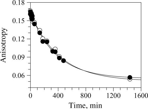
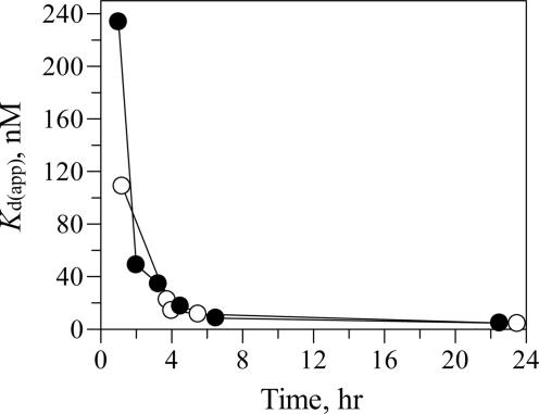
Heat shock protein (Hsp)90 is emerging as an important therapeutic target for the treatment of cancer. Two analogues of the Hsp90 inhibitor geldanamycin are currently in clinical trials. Geldanamycin (GA) and its analogues have been reported to bind purified Hsp90 with low micromolar potency, in stark contrast to their low nanomolar antiproliferative activity in cell culture and their potent antitumor activity in animal models. Several models have been proposed to account for the approximately 100-fold-greater potency in cell culture, including that GA analogues bind with greater affinity to a five-protein Hsp90 complex than to Hsp90 alone. We have determined that GA and the fluorescent analogue BODIPY-GA (BDGA) both demonstrate slow, tight binding to purified Hsp90. BDGA, used to characterize the kinetics of ligand-Hsp90 interactions, was found to bind Hsp90alpha with k(off) = 2.5 x 10(-3) min(-1), t(1/2) = 4.6 h, and Ki* = 10 nM. It was found that BDGA binds to a functional multiprotein Hsp90 complex with kinetics and affinity identical to that of Hsp90 alone. Also, BDGA binds to Hsp90 from multiple cell lysates in a time-dependent manner with similar kinetics. Therefore, our results indicate that the high potency of GA in cell culture and in vivo can be accounted for by its time-dependent, tight binding to Hsp90 alone. In the broader context, these studies highlight the essentiality of detailed biochemical characterization of drug-target interactions for the effective translation of in vitro pharmacology to cellular and in vivo efficacy.


Conflict of interest statement

Conflict of interest statement: No conflicts declared.

Figures

Fig. 1.
Fig. 1.
SDS/PAGE of the purified proteins Hsp90, Hsp70, Hsp40, Hop, and p23. Resolution of purified protein by 4–12% Bis-Tris SDS/PAGE in Mops buffer run at 200 V for 50 min. Protein loading: 2 μg per lane. Lane 1, molecular mass markers; lane 2, Hsp90α; lane 3, Hsp90β; lane 4, Hop; lane 5, Hsp70; lane 6, Hsp40; lane7, p23.
Fig. 2.
Fig. 2.
Time course of luciferase refolding in the presence of Hsp90 complex chaperone proteins. Hsp90α (2.5 μM) was incubated with 0.25 μM luciferase at 50°C for 8 min before diluting 6-fold into renaturation buffer containing 0.5 mM ATP, 2 μM Hsp70, and 1 μM Hsp40 (●). Reactions run in the absence of ATP (○), Hsp70 (□), or Hsp40 (▵) exhibit no substantial refolding activity.
Fig. 3.
Fig. 3.
Kinetic analysis of the time-dependence of BDGA binding to Hsp90α. BDGA binding to Hsp90α was measured by monitoring the fluorescence anisotropy of BDGA (10 nM) as a function of incubation time in the presence of varying concentrations of the protein. BDGA unbound in solution and bound to Hsp90α has approximate anisotropy values of 0.04 and 0.17, respectively. Data were fit to a pseudo-first-order rate equation, Eq. 8, to determine kobs values. (A) [Hsp90]: 1.5 μM (▾), 1.25 μM (▿), 0.90 μM (▴), 0.70 μM (▵), 0.50 μM (■), 0.30 μM (□), 0.10 μM (●), 0 μM (○). (B) [Hsp90]: 0.10 μM (▴), 0.075 μM (▵), 0.050 μM (■), 0.025 μM (□), 0.0125 μM (●), 0 μM (○). (C) Replot of kobs vs. [Hsp90]. Data fit to the equation for a two-step protein–ligand-binding model, Eq. 1. Kinetic constants derived from the data are summarized in Table 2.
Fig. 4.
Fig. 4.
Determination of the BDGA–Hsp90α dissociation rate (koff). BDGA was incubated with Hsp90α under conditions so that essentially all BDGA is bound to the protein. The sample is then diluted 100-fold into a solution with excess GA, and the BDGA–Hsp90α rate of dissociation measured by the time-dependent change in anisotropy under conditions where there is no appreciable BDGA–Hsp90α reassociation. ○ and ● are replicates of the same experiment. The data are fit to a monoexponential decay; average koff = 2.5 × 10⁻³ min−1, average t1/2 = 4.5 h (n = 2).
Fig. 5.
Fig. 5.
Measurement of BDGA binding to Hsp90 from cell lysates. BDGA was incubated in the presence of varying amounts of cell lysate for 1–24 h at room temperature, followed by measurement of fluorescence anisotropy. Kd(app) values were determined for BDGA–Hsp90 binding from the titration data of fluorescence anisotropy vs. [cell lysate] by using the integrated rate equation, Eq. 2. (○) Lysate from proliferating human umbilical vein endothelial cells (HUV-EC); (●) lysate from SKOV-3 cells.

References

    1. Pratt W. B. Proc. Soc. Exp. Biol. Med. 1998;217:420–434. - PubMed
    1. Pearl L. H., Prodromou C. Curr. Opin. Struct. Biol. 2000;10:46–51. - PubMed
    1. Young J. C., Hartl F. U. EMBO J. 2000;19:5930–5940. - PMC - PubMed
    1. Kosano H., Stensgard B., Charlesworth M. C., McMahon N., Toft D. J. Biol. Chem. 1998;273:32973–32979. - PubMed
    1. Isaacs J. S. Expert. Opin. Investig. Drugs. 2005;14:569–589. - PubMed

MeSH terms