. 2006 May 16;103(20):7741-6.
doi: 10.1073/pnas.0600494103. Epub 2006 May 9.

Allelic silencing at the tumor-suppressor locus 13q14.3 suggests an epigenetic tumor-suppressor mechanism

Affiliations

Allelic silencing at the tumor-suppressor locus 13q14.3 suggests an epigenetic tumor-suppressor mechanism

Daniel Mertens et al. Proc Natl Acad Sci U S A. .

Abstract

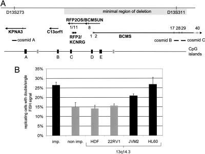
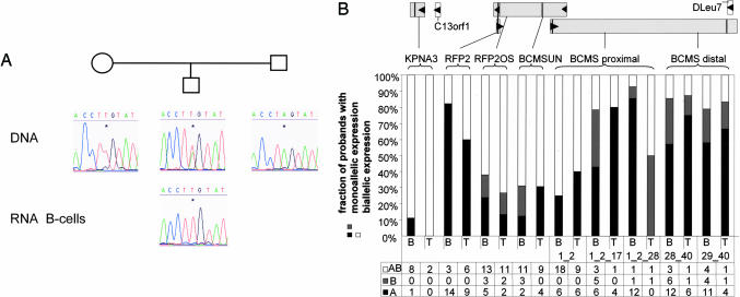
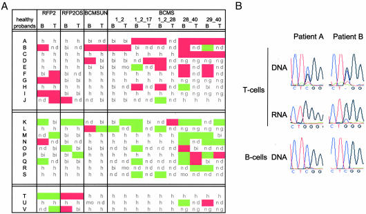
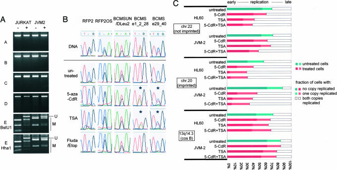
Genomic material from chromosome band 13q14.3 distal to the retinoblastoma locus is recurrently lost in a variety of human neoplasms, indicating an as-yet-unidentified tumor-suppressor mechanism. No pathogenic mutations have been found in the minimally deleted region until now. However, in B cell chronic lymphocytic leukemia tumors with loss of one copy of the critical region, respective candidate tumor-suppressor genes are down-regulated by a factor >2, which would be expected by a normal gene-dosage effect. This finding points to an epigenetic pathomechanism. We find that the two copies of the critical region replicate asynchronously, suggesting differential chromatin packaging of the two copies of 13q14.3. Although we also detect monoallelic silencing of genes localized in the critical region, monoallelic expression originates from either the maternal or paternal copy, excluding an imprinting mechanism. DNA methylation analyses revealed one CpG island of the region to be methylated. DNA demethylation of this CpG island and global histone hyperacetylation induced biallelic expression, whereas replication timing was not affected. We propose that differential replication timing represents an early epigenetic mark that distinguishes the two copies of 13q14.3, resulting in differential chromatin packaging and monoallelic expression. Accordingly, deletion of the single active copy of 13q14.3 results in significant down-regulation of the candidate genes and loss of function, providing a model for the interaction of genetic lesions and epigenetic silencing at 13q14.3 in B cell chronic lymphocytic leukemia.


Conflict of interest statement

Conflict of interest statement: No conflicts declared.

Figures

Fig. 1.
Fig. 1.
The critical region in chromosomal band 13q14.3 replicates asynchronously in cell lines. (A) Overview of the critical region between the genomic markers D13S273 and D13S311. The minimal region of deletion is shown as a long gray box. Genes and their direction of transcription are depicted as arrows. Perpendicular lines mark exons (BCMS and BCMSUN/DLEU2/RFP2OS) or SNPs (other genes) that were analyzed. Cosmids are delineated as horizontal bars. CpG islands identified in the region applying stringent conditions (24) are shown as black boxes when they are associated with 5′ ends of genes and as gray boxes when they are not. (B) Three cosmids localized in 13q14.3 (A, B, and C in A) were hybridized to interphase nuclei and compared with genomic probes localizing to an imprinted locus (imp.) and a nonimprinted locus (non imp.). Genomic probes were hybridized to nuclei of hematopoietic cell lines (HL60 and JVM-2) and nonhematopoietic cells (HDF and 22RV1). For each probe and cell line, 200 BrdU-positive cells were screened for asynchronously replicating cells with one single and one double signal (n = 4,000 cells) (for representative fluorescence in situ hybridization images, see Fig. 7). The proportion of cells showing asynchronous replication timing is depicted. Error bars give averages of the four cell lines tested (control loci) or of cosmids A, B, and C, which localize in 13q14.3.
Fig. 2.
Fig. 2.
Candidate tumor-suppressor genes in the critical region of chromosomal band 13q14.3 are monoallelically expressed in healthy probands. (A) Cells from probands heterozygous for at least one SNP were tested for monoallelic expression of candidate genes. In addition, parents of heterozygous probands were genotyped to assess the origin of the expressed allele. (B) Five genes (KPNA3, RFP2, RFP2OS, BCMSUN, and BCMS) were tested for monoallelic expression in sorted B or T cells of healthy probands. (Upper) Genes analyzed are depicted as gray boxes, arrows show the direction of transcription, and vertical lines within the genes denote the position of SNPs. For RFP2 and RFP2OS, the same SNP in exon 1L of RFP2 and exon 1 of RFP2OS was used. For BCMS, two SNPs in exons 1 and 40 were used for all splicing variants containing exons 1 and 2; 1, 2, and 17; 1, 2, and 28; 28 and 40; or 29 and 40, which are the most commonly transcribed variants of BCMS (10). (Lower) Sorted B and T cells (B and T, respectively) from probands heterozygous for at least one SNP were analyzed. Shown are percentages and absolute numbers of probands expressing both alleles (AB), the more frequently expressed allele (A), or the less frequently expressed allele (B).
Fig. 3.
Fig. 3.
Monoallelically expressed candidate genes are not imprinted but expressed from the same chromosome copy, and this active copy is deleted in CLL. (A) Parents of probands heterozygous for at least two of the tested SNPs were genotyped, and the monoallelically expressed allele of the offspring was scored for parental origin (maternal expression, green; paternal expression, red). Also shown are biallelically expressed genes (bi), monoallelically expressed genes for which parents were not available or not informative (mo), loci where the proband was homozygous (h), loci where gene expression was below the detection limit (nd), and loci where the proband was not genotyped (ng). (B) Peripheral blood lymphocytes of two CLL patients with monoallelic deletion of 13q14.3, which were informative for a SNP in RFP2OS, were sorted into a CD19-positive fraction containing mostly malignant cells (B cells) and a CD19-negative fraction containing mostly nonmalignant cells (T cells). The copy of RFP2OS active in the nonmalignant fraction is deleted in the malignant cells.
Fig. 4.
Fig. 4.
Only one CpG island in the critical region is DNA-methylated, and euchromatinization results in reexpression of the silent gene copy but no change in replication timing. (A) By using combined bisulfite and restriction analysis, DNA methylation was analyzed in CpG islands localized in 13q14.3. DNA methylation was detected in only one of five CpG islands in the critical region (see CpG island E in Fig. 1A). Upon treatment with 5-aza-CdR, DNA methylation was reduced. −, untreated cells; +, cells treated with 5-aza-CdR; U, bands representing unmethylated DNA; M, bands representing methylated DNA. (B) Different cell lines heterozygous for at least one SNP were incubated with 5-aza-CdR or TSA for 24 h and analyzed for monoallelic or biallelic expression. To rule out effects due to apoptosis, cells were also incubated with either fludarabine or etoposide. Whereas RFP2, RFP2OS, and BCMSUN did not change expression upon euchromatization in one of one and three of three cell lines tested, monoallelic silencing of the distal exons of BCMS was reduced and expression occurred from both gene copies in two of three cell lines tested. Shown are representative results of RFP2 and RFP2OS in Jurkat, BCMSUN in JVM-2, and BCMS in JVM-2 and Jurkat. (C) JVM2 and HL60 cells were treated with 5-aza-CdR or TSA, and replicating cells were scored for two unreplicated chromatids (SS), one replicated and one unreplicated chromatid (SD), or a completely replicated locus (DD). Two control loci from a nonimprinted (chromosome 22q12, 35.5 Mbp) and an imprinted region (NNAT, chromosome 20q11.2, 35.3 Mbp) shift to earlier replication after treatment with 5-aza-CdR and TSA due to euchromatization. In contrast, replication timing of 13q14.3 did not change after treatment with 5-aza-CdR or TSA.

References

    1. Liu Y., Szekely L., Grander D., Soderhall S., Juliusson G., Gahrton G., Linder S., Einhorn S. Proc. Natl. Acad. Sci. USA. 1993;90:8697–8701. - PMC - PubMed
    1. Stilgenbauer S., Nickolenko J., Wilhelm J., Wolf S., Weitz S., Döhner K., Boehm T., Döhner H., Lichter P. Oncogene. 1998;16:1891–1897. - PubMed
    1. Corcoran M. M., Hammarsund M., Zhu C., Lerner M., Kapanadze B., Wilson B., Larsson C., Forsberg L., Ibbotson R. E., Einhorn S., et al. Genes Chromosomes Cancer. 2004;40:285–297. - PubMed
    1. Liu Y., Corcoran M., Rasool O., Ivanova G., Ibbotson R., Grander D., Iyengar A., Baranova A., Kashuba V., Merup M., et al. Oncogene. 1997;15:2463–2473. - PubMed
    1. Migliazza A., Bosch F., Komatsu H., Cayanis E., Martinotti S., Toniato E., Guccione E., Qu X., Chien M., Murty V. V., et al. Blood. 2001;97:2098–2104. - PubMed

Publication types

LinkOut - more resources