. 2006 May 16;103(20):7729-34.
doi: 10.1073/pnas.0600206103. Epub 2006 May 9.

Tbx1 haploinsufficiency is linked to behavioral disorders in mice and humans: implications for 22q11 deletion syndrome

Affiliations

Tbx1 haploinsufficiency is linked to behavioral disorders in mice and humans: implications for 22q11 deletion syndrome

Richard Paylor et al. Proc Natl Acad Sci U S A. .

Abstract

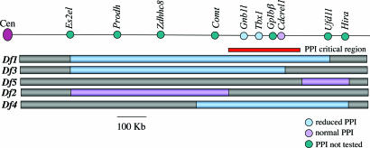
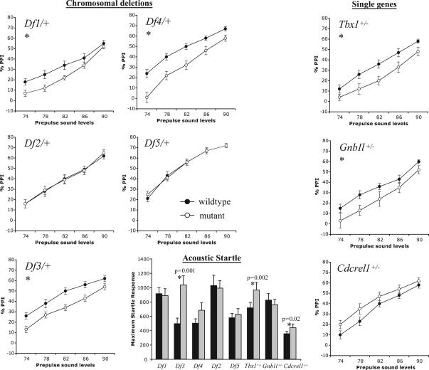
About 35% of patients with 22q11 deletion syndrome (22q11DS), which includes DiGeorge and velocardiofacial syndromes, develops psychiatric disorders, mainly schizophrenia and bipolar disorder. We previously reported that mice carrying a multigene deletion (Df1) that models 22q11DS have reduced prepulse inhibition (PPI), a behavioral abnormality and schizophrenia endophenotype. Impaired PPI is associated with several psychiatric disorders, including those that occur in 22q11DS, and recently, reduced PPI was reported in children with 22q11DS. Here, we have mapped PPI deficits in a panel of mouse mutants that carry deletions that partially overlap with Df1 and have defined a PPI critical region encompassing four genes. We then used single-gene mutants to identify the causative genes. We show that PPI deficits in Df1/+ mice are caused by haploinsufficiency of two genes, Tbx1 and Gnb1l. Mutation of either gene is sufficient to cause reduced PPI. Tbx1 is a transcription factor, the mutation of which is sufficient to cause most of the physical features of 22q11DS, but the gene had not been previously associated with the behavioral/psychiatric phenotype. A likely role for Tbx1 haploinsufficiency in psychiatric disease is further suggested by the identification of a family in which the phenotypic features of 22q11DS, including psychiatric disorders, segregate with an inactivating mutation of TBX1. One family member has Asperger syndrome, an autistic spectrum disorder that is associated with reduced PPI. Thus, Tbx1 and Gnb1l are strong candidates for psychiatric disease in 22q11DS patients and candidate susceptibility genes for psychiatric disease in the wider population.


Conflict of interest statement

Conflict of interest statement: No conflicts declared.

Figures

Fig. 1.
Fig. 1.
Deletion mutants and single-gene mutants. The black line at top is a scaled diagram of the mouse chromosome 16 region that is syntenic to human chromosome 22q11.2 showing selected genes. Blue and violet bars below indicate the deletion alleles established in mice. The original nomenclature of these alleles, Df (16)1–Df (16)5 (13, 14), has been abbreviated to Df1–Df5. The red bar indicates the PPI critical region as defined by the deletion mutants. The presence or absence of PPI impairments in mouse mutants is indicated by color, where blue indicates reduced PPI, violet indicates normal PPI, and green indicates PPI not tested.
Fig. 2.
Fig. 2.
PPI of the acoustic startle response (ASR). Reduced PPI was seen in deletion mutants Df1/+, Df3/+, and Df4/+ and in Tbx1+/− and Gnb1l+/− mice. Impairment was most apparent at the lower prepulse sound levels, as previously noted (9). The increased PPI seen in Cdcrel1+/− mice did not reach statistical significance. The magnitude of the ASR was significantly greater (∗) in Df3/+, Tbx1+/−, and Cdcrel1+/− mice than in their respective wild-type littermates, but overall there was no relationship between ASR and PPI.
Fig. 3.
Fig. 3.
Brain expression of candidate behavioral genes. Tbx1 brain expression increases steadily between E17.5 and 12 weeks as measured by real-time quantitative RT-PCR (A) and by semiquantitative RT-PCR (B). β-Gal staining of a Tbx1+/− embryo at E18.5 (C) reveals expression in blood vessels both on the brain surface (Left and Center) and within the brain parenchyma (Left). Tbx1 is expressed in the endothelial cells lining of blood vessels (arrow in Right) but not in the vascular smooth muscle (arrowheads in Right). Gnb1l expression remains steady at the same developmental stages (D). (E) β-Gal-stained thick brain sections (coronal) of an adult Gnb1l+/− mouse showing Gnb1l expression. bg, basal ganglia; t, thalamus; h, hypothalamus; p, pons; a, amygdala; c, cerebral cortex; hc, hippocampus.
Fig. 4.
Fig. 4.
Patient data and mutation analysis. (A) Family of index patient V39/02: Individuals with VCFS are shaded; circles symbolize females; squares symbolize males; and crossed symbols represent deceased family members. ?, no information available. (B) Shown at the top is a schematic representation of alternatively spliced TBX1 transcripts (TBX1A, TBX1B, TBX1C); arrows indicate position of known frameshift mutations in TBX1C; green boxes depict TBX1 coding sequences; and gray boxes depict UTRs. Shown at the bottom is a DNA sequence of a patient with the 1320–1342del23bp mutation (upper panel) and the wild-type TBX1 sequence in an unrelated individual (lower panel); the position of the mutation is boxed. (C) Subcellular localization of wild-type and mutant TBX1 constructs expressed in U2-OS cells. hTbx1, wild-type TBX1; 1320–1342del, del23bp mutation described here; G145R, predicted null mutation; 1250delC, point mutation described by Yagi et al. (26). Constructs were detected with anti-TBX1 antibody. Cell nuclei were stained with DAPI. (D) Transcriptional activation of the CAT reporter gene by wild-type and mutant TBX1. Significant differences in transcriptional activation between wild-type TBX1 and a TBX1 construct are indicated by ∗∗∗ (P ≤ 0.001). Data were normalized for transfection efficiency and depicted as average values ± SEM.

References

    1. Fine S. E., Weissman A., Gerdes M., Pinto-Martin J., Zackai E. H., McDonald-McGinn D. M., Emanuel B. S. J. Autism Dev. Disord. 2005;35:461–470. - PMC - PubMed
    1. Niklasson L., Rasmussen P., Oskarsdottir S., Gillberg C. Genet. Med. 2001;3:79–84. - PubMed
    1. Bassett A. S., Hodgkinson K., Chow E. W., Correia S., Scutt L. E., Weksberg R. Am. J. Med. Genet. 1998;81:328–337. - PMC - PubMed
    1. Murphy K. C., Jones L. A., Owen M. J. Arch. Gen. Psychiatry. 1999;56:940–945. - PubMed
    1. Bassett A. S., Chow E. W., AbdelMalik P., Gheorghiu M., Husted J., Weksberg R. Am. J. Psychiatry. 2003;160:1580–1586. - PMC - PubMed

Publication types

MeSH terms

Substances