. 2006 May 16;103(20):7682-7.
doi: 10.1073/pnas.0600148103. Epub 2006 May 9.

siRNA in human cells selectively localizes to target RNA sites

Affiliations

siRNA in human cells selectively localizes to target RNA sites

Svitlana Y Berezhna et al. Proc Natl Acad Sci U S A. .

Abstract

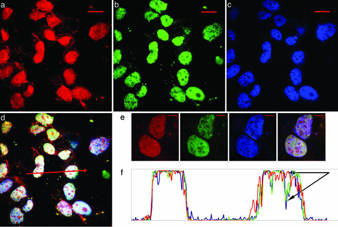
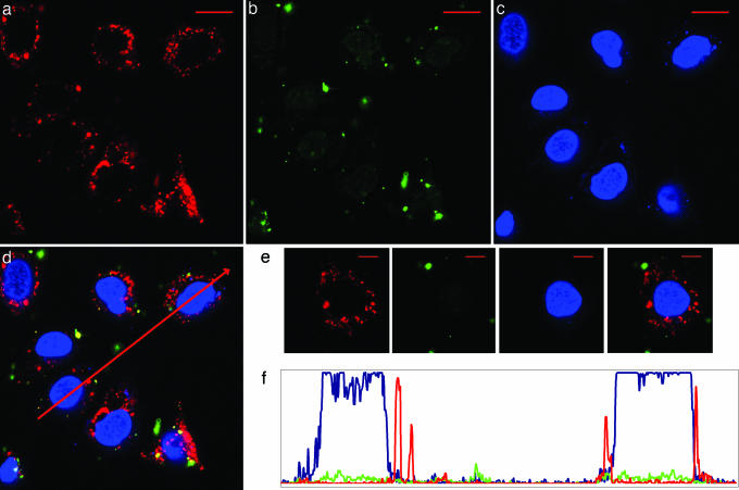
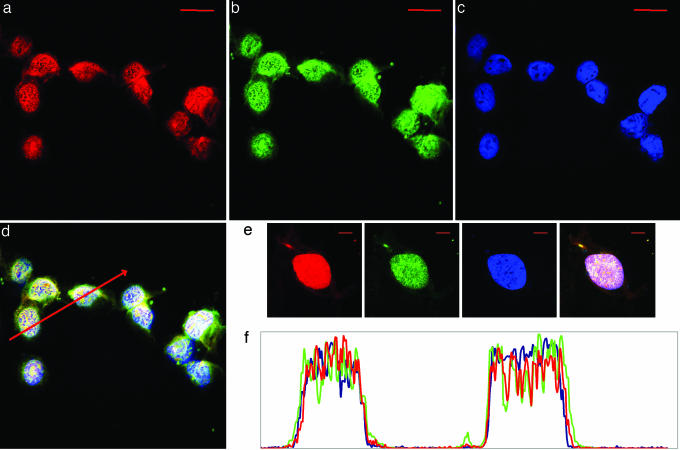
Recent observations of RNA interference (RNAi) in the nuclei of human cells raise key questions about the extent to which nuclear and cytoplasmic RNAi pathways are shared. By directly visualizing the localization of small interfering RNA (siRNA) in live human cells, we show here that siRNA either selectively localizes in the cytoplasm or translocates into the nucleus, depending on where the silencing target RNA resides. Two siRNAs that target the small nuclear 7SK and U6 RNAs localize into the nucleus as duplexes. In contrast, an siRNA targeting the cytoplasmic hepatitis C virus replicon RNA dissociates, and only antisense strand distributes in the cytoplasm of the cells harboring the target RNA, whereas sense strand gets degraded. At the same time, both strands of the latter siRNA are distributed throughout the cytoplasm and nucleus in cells lacking the silencing target RNA. These results suggest the existence of a mechanism by which the RNAi machinery orchestrates a target-determined localization of the siRNA and the corresponding RNAi activity, and also provide evidence for formation of nuclear-programmed active RNA induced silencing complexes directly in the nucleus.


Conflict of interest statement

Conflict of interest statement: No conflicts declared.

Figures

Fig. 1.
Fig. 1.
7SK siRNA accumulates in the nucleus in Huh-7 cells 2 h after transfection. (ac) 7SK siRNA antisense (red) and sense (green) strands colocalize primarily in the nucleus (blue), seen in the cytoplasm only in small amounts. (Scale bar, 20 μm.) (d) Overlay of red, green, and blue. (e) Two cells. (Scale bar, 10 μm.) (f) Intensity profiles along the selected line (red arrow in d) across cells show that both siRNA strands enter the nucleus, where they apparently get separated, because primarily the antisense strand is visible in the nucleoli (marked by arrows, DAPI-depleted dark spots against a blue background in c).
Fig. 2.
Fig. 2.
NS3 siRNA appears restricted to the cytoplasm of Huh-7 cells 2 h after transfection. (ac) NS3 siRNA antisense strand (red) is seen clustered in the perinuclear area but does not enter the nucleus (blue), whereas sense strand (green) is barely visible. (Scale bar, 20 μm.) (d) Overlay of red, green, and blue. (e) A single cell. (Scale bar, 10 μm.) (f) Intensity profiles along the selected line (red arrow in d) across cells confirm perinuclear antisense strand (red) localization and show that a small amount of sense (green) strand can be found within the nucleus (blue).
Fig. 3.
Fig. 3.
NS3 siRNA distributes into the cytoplasm as well as the nucleus in CHO cells 2 h after transfection. (ac) NS3 siRNA antisense (red) and sense (green) strands are seen throughout the cytoplasm and the nucleus (blue). (Scale bar, 20 μm.) (d) Overlay of red, green, and blue. (e) A single cell. (Scale bar, 10 μm.) (f) Intensity profiles along the selected line (red arrow in d) across two cells.
Fig. 4.
Fig. 4.
7SK siRNA localizes primarily to the nucleus in CHO cells 2 h after transfection. (ac) 7SK siRNA antisense (red) and sense (green) strands accumulate in the nucleus (blue), showing very low cytoplasmic distribution. (Scale bar, 20 μm.) (d) Overlay of red, green, and blue. (e) A single cell. (Scale bar, 10 μm.) (f) Intensity profiles along the selected line.
Fig. 5.
Fig. 5.
7SK siRNA distributes throughout CHO cells 24 h after transfection. (ac) 7SK siRNA antisense strand (red) is seen in the cytoplasm and the nucleus, whereas sense strand (green) gets eliminated. (d) Colocalization shows antisense strand is retained in the nucleoli (red spots) and confirms integrity of the cell nuclei (blue). (Scale bar, 20 μm.) (e) A single cell. (Scale bar, 10 μm.) (f) Intensity profiles along the selected line.

References

    1. Fire A., Xu S., Montgomery M. K., Kostas S. A., Driver S. E., Mello C. C. Nature. 1998;391:806–811. - PubMed
    1. Morris K. V., Chan S. W., Jacobsen S. E., Looney D. J. Science. 2004;305:1289–1292. - PubMed
    1. Kawasaki H., Taira K. Nature. 2004;431:211–217. - PubMed
    1. Langlois M. A., Boniface C., Wang G., Alluin J., Salvaterra P. M., Puymirat J., Rossi J. J., Lee N. S. J. Biol. Chem. 2005;280:16949–16954. - PubMed
    1. Haussecker D., Proudfoot N. J. Mol. Cell. Biol. 2005;25:9724–9733. - PMC - PubMed

Publication types