. 2006 Aug 15;398(1):113-24.
doi: 10.1042/BJ20060280.

Kinetic and chemical analyses of the cytokinin dehydrogenase-catalysed reaction: correlations with the crystal structure

Affiliations

Kinetic and chemical analyses of the cytokinin dehydrogenase-catalysed reaction: correlations with the crystal structure

Hana Popelková et al. Biochem J. .

Abstract

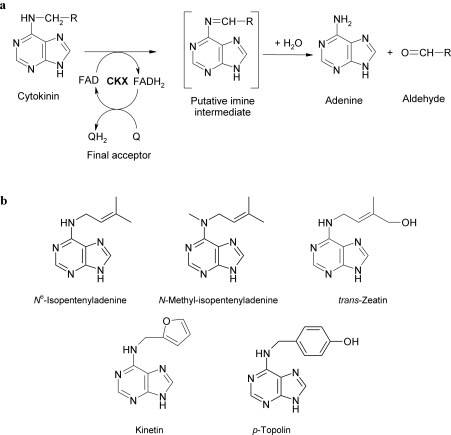
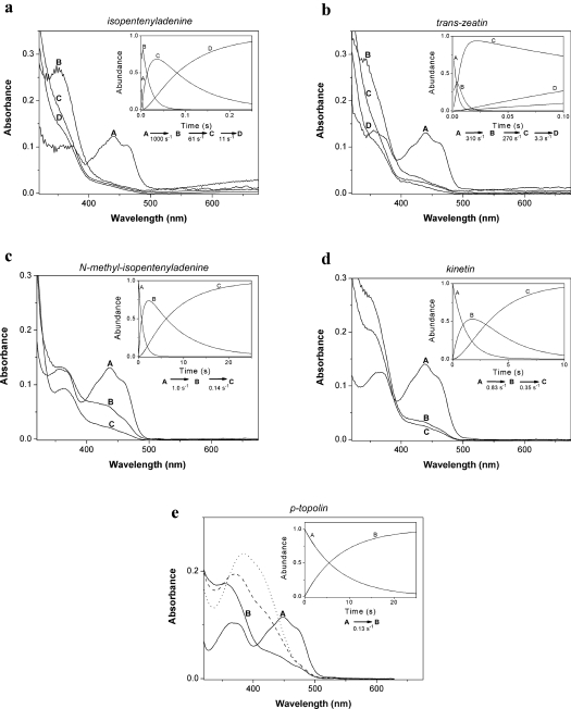
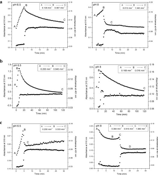
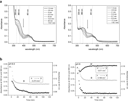
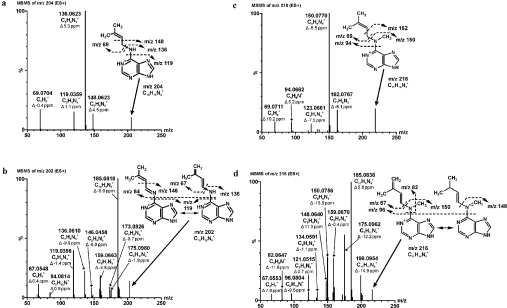
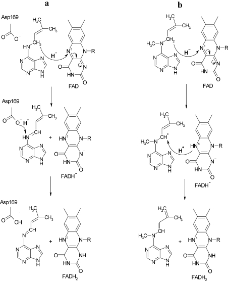
CKX (cytokinin dehydrogenase) is a flavoprotein that cleaves cytokinins to adenine and the corresponding side-chain aldehyde using a quinone-type electron acceptor. In the present study, reactions of maize (Zea mays) CKX with five different substrates (N6-isopentenyladenine, trans-zeatin, kinetin, p-topolin and N-methyl-isopentenyladenine) were studied. By using stopped-flow analysis of the reductive half-reaction, spectral intermediates were observed indicative of the transient formation of a binary enzyme-product complex between the cytokinin imine and the reduced enzyme. The reduction rate was high for isoprenoid cytokinins that showed formation of a charge-transfer complex of reduced enzyme with bound cytokinin imine. For the other cytokinins, flavin reduction was slow and no charge-transfer intermediates were observed. The binary complex of reduced enzyme and imine product intermediate decays relatively slowly to form an unbound product, cytokinin imine, which accumulates in the reaction mixture. The imine product only very slowly hydrolyses to adenine and an aldehyde derived from the cytokinin N6 side-chain. Mixing of the substrate-reduced enzyme with Cu2+/imidazole as an electron acceptor to monitor the oxidative half-reaction revealed a high rate of electron transfer for this type of electron acceptor when using N6-isopentenyladenine. The stability of the cytokinin imine products allowed their fragmentation analysis and structure assessment by Q-TOF (quadrupole-time-of-flight) MS/MS. Correlations of the kinetic data with the known crystal structure are discussed for reactions with different cytokinins.


Figures

Figure 1
Figure 1. Cytokinin dehydrogenase and cytokinins
(a) Reaction mechanisms of cytokinin dehydrogenase (EC 1.5.99.12), (b) structures of cytokinins used in the present study.
Figure 2
Figure 2. Anaerobic reduction of ZmCKX1 with cytokinins
Using a stopped-flow instrument equipped with a photodiode array detector, spectral data were obtained upon anaerobic mixing of ZmCKX1 with cytokinin substrates. The spectral species shown were derived from global analysis of the data obtained. The data could be fitted using a model in which species A decays in an exponential fashion to form B (A→B). If needed, more exponential decay steps were included in the model. Typically, 23 μM ZmCKX1 was mixed with 0.5 mM of a cytokinin substrate (except for N-methyl-isopentenyladenine and kinetin where 0.25 mM was used) under anaerobic conditions in 75 mM imidazole/HCl (pH 6.5) at 25 °C. The deconvoluted spectra are shown for: (a) isopentenyladenine, (b) trans-zeatin, (c) N-methyl-isopentenyladenine, (d) kinetin, and (e) p-topolin. For p-topolin (e), spectra of species B obtained with 75 mM Tris/HCl (pH 7.5; broken line) and (pH 8.5; dotted line) are shown. Time courses obtained by global analyses for the respective species are shown in the insets. For experimental details see the Materials and methods section.
Figure 3
Figure 3. Cytokinin intermediate formation and decay
Time courses of the reaction intermediate formation and decay (310 nm; circles) and oxidation changes of the FAD cofactor (451 nm; triangles) observed for ZmCKX1 (5 μM) that was mixed aerobically with 125 μM cytokinins [(a) isopentenyladenine, (b) N-methyl-isopentenyladenine, and (c) trans-zeatin] in 75 mM imidazole/HCl (pH 6.5; left-hand panels) and 75 mM Tris/HCl (pH 8.0; right-hand panels). The data could be fitted using a model in which species A is converted in a hyperbolic fashion into species B that then decays exponentially to form C.
Figure 4
Figure 4. Interaction of ZmCKX1 with p-topolin
(a) Spectral changes in the range of 250–700 nm, and (b) time courses of the reaction intermediate decay (360 nm, open circles), reduction of the FAD cofactor (451 nm, triangles) and formation of the product 4-hydroxybenzaldehyde (330 nm at pH 8.0, filled circles) observed for ZmCKX1 (5 μM) that was mixed aerobically with 125 μM p-topolin in 75 mM imidazole/HCl (pH 6.5; left-hand panels) and with 13 μM p-topolin in 75 mM Tris/HCl (pH 8.0; right-hand panels). Arrows in (a) indicate the direction of spectral changes as observed from 1.3 min to 118.7 min of the reaction course. The data (b) could be fitted using a model in which species A decays in an exponential fashion to form B. To differentiate the kinetics of the formation of 4-hydroxybenzaldehyde (330 nm) at pH 8, the species are marked as A' and B'.
Figure 5
Figure 5. MS/MS analysis of the ZmCKX1 reaction with cytokinins
Cytokinins [(a) isopentenyladenine (50 μM), (b) isopentenyladenine[-2H] intermediate, (c) N-methyl-isopentenyladenine (50 μM) and (d) N-methyl-isopentenyladenine[-2H] intermediate] were dissolved in 30 mM ammonium hydrogen carbonate (pH 8.0). For the reaction intermediate analysis, a reaction mixture that contained 1.5 μM ZmCKX1 and 50 μM cytokinin in 30 mM ammonium hydrogen carbonate (pH 8.0) was prepared and incubated at 25 °C for 2 min before the measurement. The high-resolution MS/MS experiments were done on a hybrid mass analyser Q-TOF micro with MassLynx data system software. The samples were applied in the form of an in-line spray at an injection rate of 10 μl/min. The data acquisition was performed in the range 50–1000 Da with a cycle time 47 μs, scan time 0.5 s, and collision energy 15, 20, 25, 30 and 35 V. The accurate masses of the parent ions and their fragments were calculated and used for the determination of the elementary composition and structure with fidelity within 15 p.p.m.
Figure 6
Figure 6. Reaction mechanisms of cytokinin dehydrogenase for (a) isopentenyladenine, as described by Malito et al. [9], and (b) N-methyl-isopentenyladenine proposed in the present study
Note that Asp169 could also act as a base, instead of the N5 of the reduced flavin (see Discussion section).

References

    1. van Berkel W. J. H., Benen J. A. E., Eppink M. H. M., Fraaije M. W. Flavoprotein kinetics. In: Chapman S. K., Reid G. A., editors. Methods in Molecular Biology, vol. 131, Flavoprotein Protocols. Totowa, NJ: Humana Press; 1999. pp. 61–86. - PubMed
    1. Hare P. D., van Staden J. Cytokinin oxidase: biochemical features and physiological significance. Physiol. Plant. 1994;91:128–136.
    1. Galuszka P., Frébort I., Šebela M., Peč P. Degradation of cytokinins by cytokinin oxidases in plants. Plant Growth Regul. 2000;32:315–327.
    1. Galuszka P., Frébort I., Šebela M., Sauer P., Jacobsen S., Peč P. Cytokinin oxidase or dehydrogenase?. Mechanism of cytokinin degradation in cereals. Eur. J. Biochem. 2001;268:450–461. - PubMed
    1. Frébortová J., Fraaije M. W., Galuszka P., Šebela M., Peč P., Hrbáč J., Novák O., Bilyeu K. D., English J. T., Frébort I. Catalytic reactions of cytokinin dehydrogenase: preferences for quinones as electron acceptors. Biochem. J. 2004;380:121–130. - PMC - PubMed

Publication types