Case Reports
. 2006 Aug 15;398(1):107-12.
doi: 10.1042/BJ20060221.

Secondary mitochondrial dysfunction in propionic aciduria: a pathogenic role for endogenous mitochondrial toxins

Affiliations
Case Reports

Secondary mitochondrial dysfunction in propionic aciduria: a pathogenic role for endogenous mitochondrial toxins

Marina A Schwab et al. Biochem J. .

Abstract

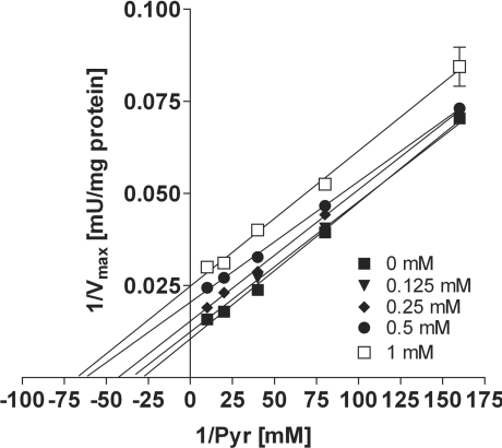
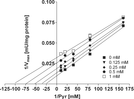
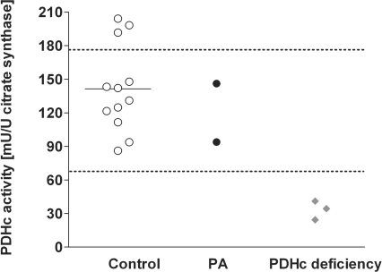
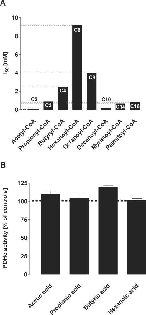
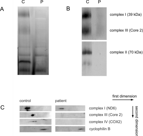
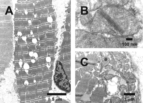
Mitochondrial dysfunction during acute metabolic crises is considered an important pathomechanism in inherited disorders of propionate metabolism, i.e. propionic and methylmalonic acidurias. Biochemically, these disorders are characterized by accumulation of propionyl-CoA and metabolites of alternative propionate oxidation. In the present study, we demonstrate uncompetitive inhibition of PDHc (pyruvate dehydrogenase complex) by propionyl-CoA in purified porcine enzyme and in submitochondrial particles from bovine heart being in the same range as the inhibition induced by acetyl-CoA, the physiological product and known inhibitor of PDHc. Evaluation of similar monocarboxylic CoA esters showed a chain-length specificity for PDHc inhibition. In contrast with CoA esters, non-esterified fatty acids did not inhibit PDHc activity. In addition to PDHc inhibition, analysis of respiratory chain and tricarboxylic acid cycle enzymes also revealed an inhibition by propionyl-CoA on respiratory chain complex III and alpha-ketoglutarate dehydrogenase complex. To test whether impairment of mitochondrial energy metabolism is involved in the pathogenesis of propionic aciduria, we performed a thorough bioenergetic analysis in muscle biopsy specimens of two patients. In line with the in vitro results, oxidative phosphorylation was severely compromised in both patients. Furthermore, expression of respiratory chain complexes I-IV and the amount of mitochondrial DNA were strongly decreased, and ultrastructural mitochondrial abnormalities were found, highlighting severe mitochondrial dysfunction. In conclusion, our results favour the hypothesis that toxic metabolites, in particular propionyl-CoA, are involved in the pathogenesis of inherited disorders of propionate metabolism, sharing mechanistic similarities with propionate toxicity in micro-organisms.


Figures

Figure 1
Figure 1. Inhibition of PDHc by propionyl-CoA
Spectrophotometric analysis in purified PDHc from porcine heart demonstrates uncompetitive inhibition of PDHc activity by propionyl-CoA with respect to pyruvate (Pyr) as demonstrated by a Lineweaver–Burk plot. Values are given as the means±S.D. (where no error bars are visible the S.D. is within the symbol). Experiments were performed at least in triplicate.
Figure 2
Figure 2. Inhibition of PDHc by acetyl-CoA
Spectrophotometric analysis in purified PDHc from porcine heart demonstrates uncompetitive inhibition of PDHc activity by acetyl-CoA with respect to pyruvate (Pyr) as demonstrated by a Lineweaver–Burk plot. Values are given as the means±S.D. (where no error bars are visible the S.D. is within the symbol). Experiments were performed at least in triplicate.
Figure 3
Figure 3. Spectrophotometric analysis of PDHc activity in human skin fibroblasts
Spectrophotometric analysis of PDHc activity was performed in human skin fibroblasts from healthy volunteers (n=12), patients with PA (n=2) and primary PDHc deficiency (n=3). The horizontal bar represents the mean for the control group and the dotted lines indicate the range of the control group.
Figure 4
Figure 4. Chain-length-specific effects of acyl-CoA esters (A) and fatty acids (B) on PDHc activity
(A) Acyl-CoA esters: spectrophotometric analysis of PDHc activity was performed in purified enzyme from porcine heart using short-, medium- and long-chain monocarboxylic acyl-CoA esters. The Figure summarizes the I50 values of acyl-CoA esters. Values are given as means. Experiments were performed at least in triplicate. (B) Fatty acids: in contrast with acyl-CoA esters, corresponding short- and medium-chain fatty acids (up to 1 mM) did not inhibit PDHc activity in purified enzyme from porcine heart. PDHc activity was determined under standard conditions. Values were normalized to 100% (dotted line) and are given as the means±S.D. Experiments were performed at least in triplicate.
Figure 5
Figure 5. BN-PAGE of OXPHOS complexes in muscle homogenates
Proteins (40 μg) of solubilized muscle homogenates from patient 2 (P) and control (C) were analysed by BN-PAGE (5–15% gel) for the separation of multisubunit complexes. (A) In-gel activity assay of mitochondrial complex I confirming a decrease in activities of patient 2 compared with control samples. (B) A second gel was run in duplicate and Western-blot analysis was performed using antibodies against complex I subunit 39 kDa, complex II subunit 70 kDa and complex III subunit Core 2. (C) A second dimension was run and Western-blot analysis was performed using antibodies against OXPHOS complexes I (ND6), III (Core 2) and IV [COX2 (cytochrome oxidase 2)] and cyclophilin B as a loading control. Arrows indicate the first and second dimensions.
Figure 6
Figure 6. Ultrastructural changes in muscle tissue of PA patients
Electron microscopy of muscle biopsy specimens (musculus vastus lateralis dexter) of both PA patients was performed. (A) Patient 1: muscle fibre with many lipid droplets (the large clear globules). (B) Patient 1: mitochondrion with a crystalline inclusion. (C) Patient 2: large swollen mitochondrion with disrupted cristae and two mitochondria with a dark globular inclusion.

References

    1. Fenton W. A., Gravel R. A., Rosenblatt D. S. Disorders of propionate and methylmalonate metabolism. In: Scriver C. R., Beaudet A. L., Valle A. D., Sly W. S., editors. The Metabolic and Molecular Bases of Inherited Disease. New York: McGraw-Hill; 2001. pp. 2165–2193.
    1. Miyazaki T., Ohura T., Kobayashi M., Shigematsu Y., Yamaguchi S., Suzuki Y., Hata I., Aoki Y., Yang X., Minjares C., et al. Fatal propionic acidemia in mice lacking propionyl-CoA carboxylase and its rescue by postnatal, liver-specific supplementation via a transgene. J. Biol. Chem. 2001;276:35995–35999. - PubMed
    1. Brismar J., Ozand P. T. CT and MR of the brain in disorders of the propionate and methylmalonate metabolism. Am. J. Neuroradiol. 1994;15:1459–1473. - PMC - PubMed
    1. Brock M., Buckel W. On the mechanism of action of the antifungal agent propionate. Eur. J. Biochem. 2004;271:3227–3241. - PubMed
    1. Cheema-Dhadli S., Leznoff C. C., Halperin M. L. Effect of 2-methylcitrate on citrate metabolism: implications for the management of patients with propionic acidemia and methylmalonic aciduria. Pediatr. Res. 1975;9:905–908. - PubMed

Publication types

MeSH terms