. 2006 May;17 Suppl 1(Suppl 1):S169-S177.
doi: 10.1111/j.1540-8167.2006.00401.x.

Ranolazine improves abnormal repolarization and contraction in left ventricular myocytes of dogs with heart failure by inhibiting late sodium current

Affiliations

Ranolazine improves abnormal repolarization and contraction in left ventricular myocytes of dogs with heart failure by inhibiting late sodium current

Albertas I Undrovinas et al. J Cardiovasc Electrophysiol. 2006 May.

Abstract

Background: Ventricular repolarization and contractile function are frequently abnormal in ventricular myocytes from human failing hearts as well as canine hearts with experimentally induced heart failure (HF). These abnormalities have been attributed to dysfunction involving various steps of the excitation-contraction coupling process, leading to impaired intracellular sodium and calcium homeostasis. We previously reported that the slow inactivating component of the Na(+) current (late I(Na)) is augmented in myocytes from failing hearts, and this appears to play a significant role in abnormal ventricular myocytes repolarization and function. We tested the effect of ranolazine, a novel drug being developed to treat angina, on (1) action potential duration (APD), (2) peak transient and late I(Na) (I(NaT) and I(NaL), respectively), (3) early afterdepolarizations (EADs), and (4) twitch contraction (TC), including after contractions and contracture.

Methods: Myocytes were isolated from the left ventricle of normal dogs and of dogs with chronic HF caused by multiple sequential intracoronary micro-embolizations. I(NaT) and I(NaL) were recorded using conventional whole-cell patch-clamp techniques. APs were recorded using the beta-escin perforated patch-clamp configuration at frequencies of 0.25 and 0.5 Hz. TCs were recorded using an edge movement detector at stimulation frequencies ranging from 0.5 to 2.0 Hz.

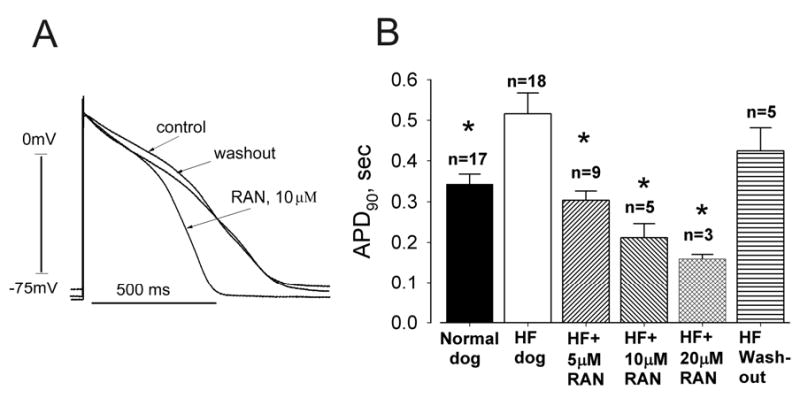
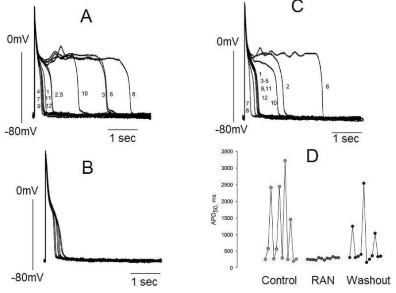
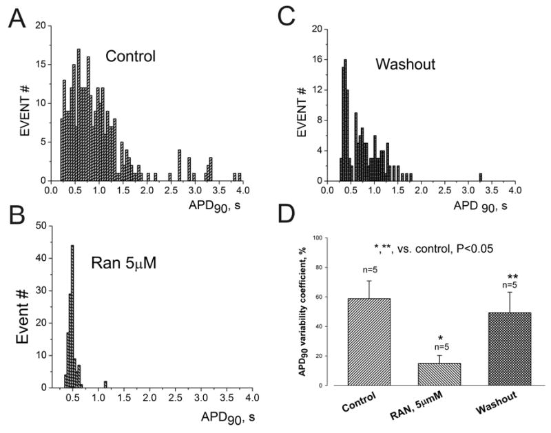
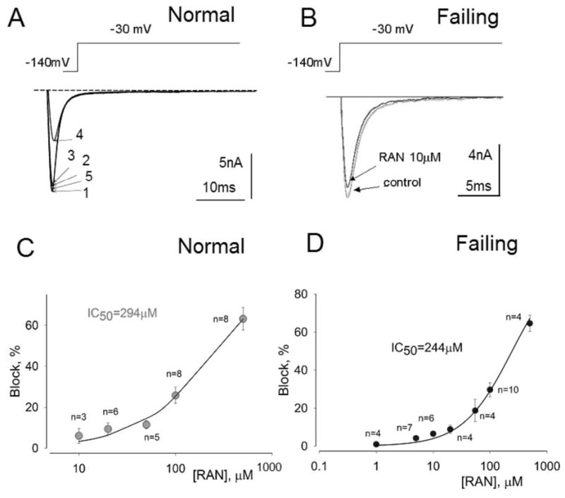
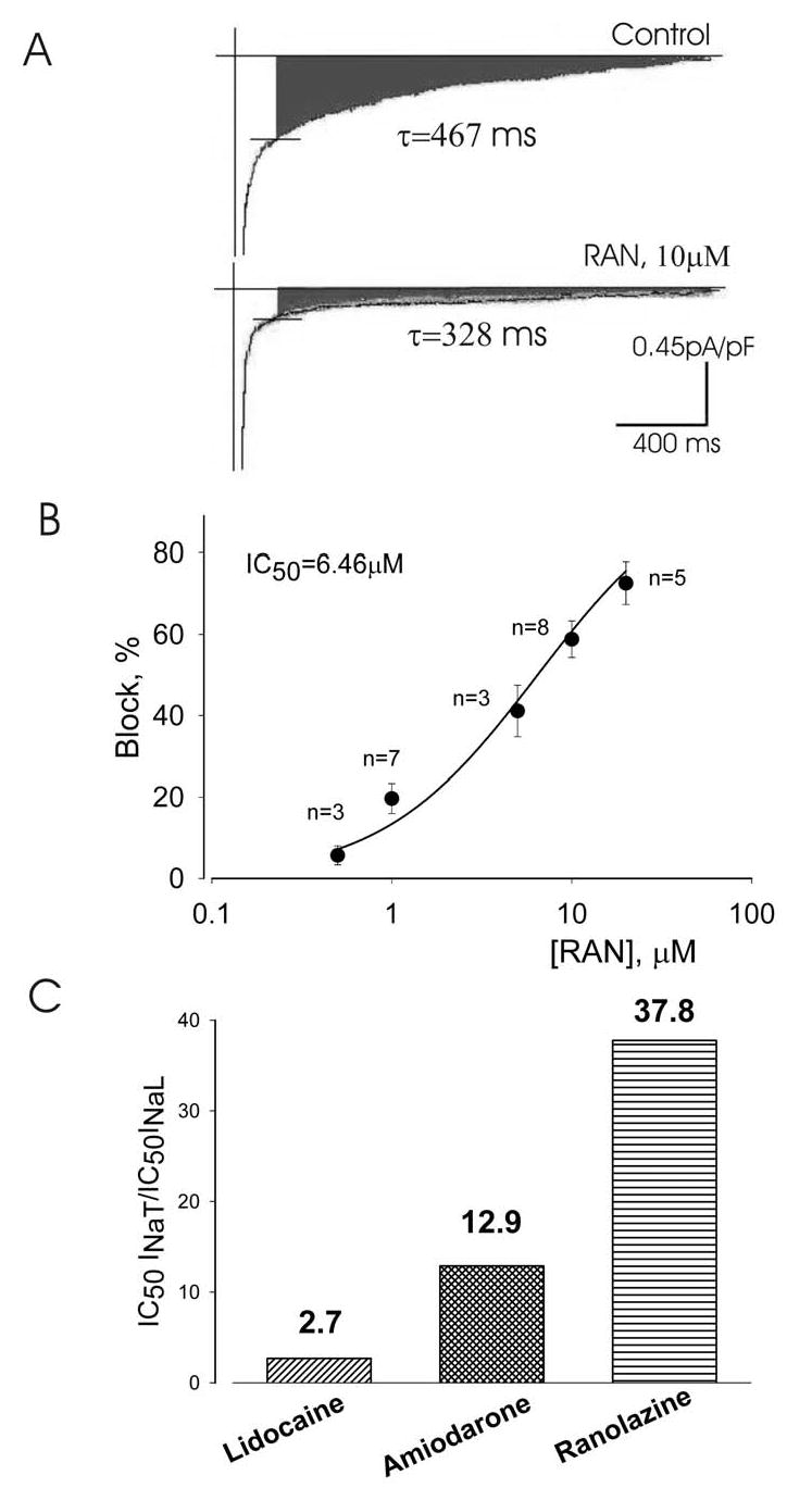
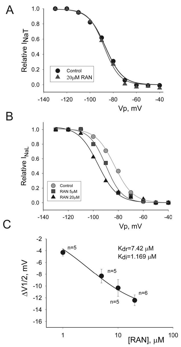
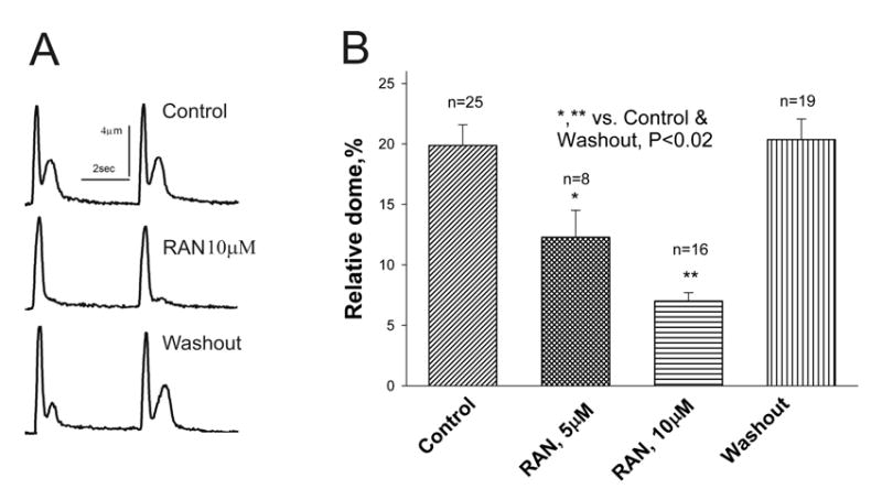
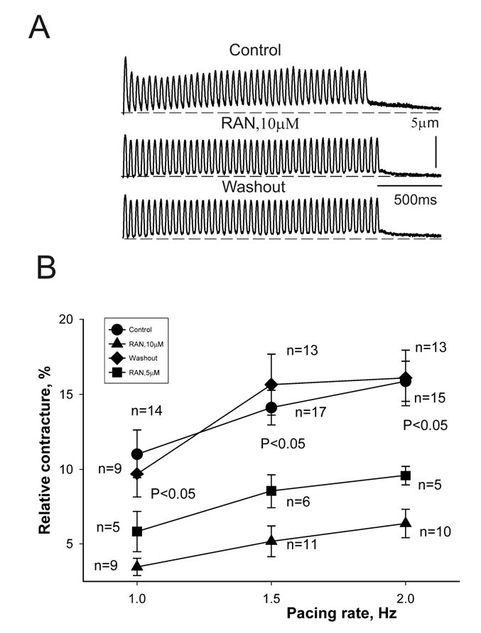
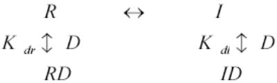
Results: Ranolazine significantly (P<0.05) and reversibly shortened the APD of myocytes stimulated at either 0.5 or 0.25 Hz in a concentration-dependent manner. At a stimulation frequency of 0.5 Hz, 5, 10, and 20 microM ranolazine shortened the APD(90) (APD measured at 90% repolarization) from 516+/-51 to 304+/-22, 212+/-34 and 160+/-11 ms, respectively, and markedly decreased beat-to-beat variability of APD(90), EADs, and dispersion of APDs. Ranolazine preferentially blocked I(NaL) relative to I(NaT) in a state-dependent manner, with a approximately 38-fold greater potency against I(NaL) to produce tonic block (IC(50)=6.5 microM) than I(NaT) (IC(50)=294 microM). When we evaluated inactivated state blockade of I(NaL) from the steady-state inactivation mid-potential shift using a theoretical model, ranolazine was found to bind more tightly to the inactivated state than the resting state of the sodium channel underlying I(NaL), with apparent dissociation constants K(dr)=7.47 microM and K(di)=1.71 microM, respectively. TCs of myocytes stimulated at 0.5 Hz were characterized by an initial spike followed by a dome-like after contraction, which was observed in 75% of myocytes from failing hearts and coincided with the long AP plateau and EADs. Ranolazine at 5 and 10 microM reversibly shortened the duration of TCs and abolished the after contraction. When the rate of myocyte stimulation was increased from 1.0 to 2.0 Hz, there was a progressive increase in diastolic "tension," that is, contracture. Ranolazine at 5 and 10 microM reversibly prevented this frequency-dependent contracture.


Figures

Figure 1
Figure 1
Ranolazine (RAN) reversibly shortens action potential (AP) duration (APD) of ventricular myocytes isolated from canine failing hearts (HF). Panel A: Representative APs of single left ventricular cardiomyocytes. Superimposed APs were recorded at a pacing rate of 0.5 Hz in the absence of drug (control), in the presence of ranolazine (10 μM) and after drug washout. Panel B: Effect of ranolazine on APD at 90% repolarization (APD90) of ventricular myocytes stimulated at a rate of 0.5 Hz. Ranolazine at concentrations of 5, 10 and 20 μM significantly and reversibly shortened APD90. Data were pooled from 3 non-failing and 6 failing hearts. All APs longer than 2 s were excluded. n= number of myocytes; * p < 0.05 vs. HF (ANOVA followed by Bonferroni’s post-hoc test against HF).
Figure 2
Figure 2
Ranolazine (RAN) reduces APD variability in left ventricular myocytes isolated from canine failing hearts. Twelve consecutive APs recorded at a pacing rate of 0.25 Hz are superimposed. Panel A: APs in the absence of drugs (control). Panel B: APs recorded in the presence of 10 μM of ranolazine. Panel C: APs recorded 3–4 minutes after drug washout. Panel D: Summary of the data shown in panels A-C. Each data point represents the APD50 from a series of APs recorded consecutively at a pacing rate of 0.25 Hz. The numbers next to the AP tracings in panels A and C are consecutive APs recorded during a period of ~ 45 seconds.
Figure 3
Figure 3
Ranolazine (RAN) reversibly reduces APD dispersion in left ventricular myocytes isolated from canine failing hearts. Histograms of APD distribution measured at 90% repolarization (APD90).APs were recorded in the absence of drugs (control) (A), in the presence of 5 μM ranolazine (B), and after drug washout (C). Bin size = 0.05 s for all histograms. D. APD variability measured as the coefficient of variability, CV = (SD/mean APD90) x 100%. Ranolazine (5 μM) reversibly reduced ADP90 variability in failing myocytes. Data were obtained in 5 ventricular myocytes from 3 failing dog hearts. P < 0.05 control vs. RAN and RAN vs. washout (ANOVA).
Figure 4
Figure 4
Ranolazine (RAN) weakly inhibits peak transient sodium current. Effect of ranolazine on peak INa recorded from left ventricular myocytes isolated from normal (A, C) and failing canine hearts (B,D). Panel A: Superimposed current traces recorded in a non failing myocyte in the absence of drugs (1), and in the presence of 10 (2), 20 (3) and 500 μM ranolazine (4) and after drug washout (5). Panel B: Superimposed current traces recorded without (control) and with 10 μM ranolazine recorded in failing myocytes. Panels C and D: Concentration-response relationships for inhibition of peak INa by ranolazine in normal (C) and failing hearts (D). Points and error bars represent mean and standard error of peak sodium currents obtained in 13 myocytes from 2 normal (non-failing) hearts (C) and 14 myocytes from 4 failing hearts (D). Data points are fitted to a one-to-one binding model (solid line; Equation 1, Methods). The IC50 value is the concentration of ranolazine that caused 50% inhibition of peak INa.
Figure 5
Figure 5
Inhibition by ranolazine (RAN) of the late INa of left ventricular myocytes isolated from canine failing hearts. Panel A: Traces of late INa recorded without (control), and with 10 μM ranolazine. The peak transient inward sodium current is truncated. The decay time constant for late INa (τ values, single exponential fit; Equation 4, Methods) is given next to the current traces. Panel B: Concentration-response relationship for the inhibition of late INa by ranolazine. Points represent from 6 myocytes from 2 hearts. Data points are fit to a one-to-one binding model (Equation 1, Methods). The amplitude of late INaL was determined as the average current within 200–220 ms after the onset of a 2-s depolarization. Data are mean ± SEM. Panel C: Potencies of lidocaine, amiodarone and ranolazine to inhibit peak and late INa representing the drug concentrations necessary to cause 50% block of peak and late INa.
Figure 6
Figure 6
Concentration-dependent effect of ranolazine (RAN) on steady-state inactivation (SSI) of peak transient INa (INaT) and late INa (INaL) recorded in left ventricular myocytes isolated from canine failing hearts. Panel A: SSI-voltage relationship for peak INa in controls (•), and in the presence of 20 μM of ranolazine (▴), which failed to shift the SSI curve. Solid lines in Panels A and B represent data point fit to the Boltzmann function (Equation 2, Methods). All data were obtained in the same cell. Panel B: SSI-voltage relationship for late INa in controls (•) and in the presence of 5 (▪), and 10 μM ranolazine (▴), showing the leftward shift of the SSI mid-potential caused by ranolazine. All data were obtained in the same cell. Panel C: SSI mid potential shift (ΔV½) caused by ranolazine in 11 ventricular myocytes from 5 failing hearts. Solid line represents a data fit to equation 3 (Methods) to obtain dissociation constants of ranolazine for the resting (Kdr) and inactivated (Kdi) states of INaL as shown in the graph. The slope factor k in equation 3 was taken as 7.6mV, the evaluated mean for the cells presented in the panel C. Data are means ± SEM.
Figure 7
Figure 7
Ranolazine inhibition of abnormal relaxation of twitch contractions representing single ventricular myocytes from failing dog hearts. Panel A: Individual twitch shortenings recorded in control conditions (no drug, top), in the presence of ranolazine (10 μM, middle), and after drug washout (bottom). Panel B: Data from myocytes of 6 dogs with HF. Twitch contraction of a failing myocyte is characterized by a spike-dome shape. Ranolazine normalized the shape of the twitch contraction by abolishing the dome (tonic) component. *p < 0.001, **p < 0.02 vs. control (ANOVA).
Figure 8
Figure 8
Ranolazine inhibition of frequency-dependent contracture (i.e., increase in diastolic tension) in failing myocytes. Panel A: Twitch contractions recorded at a pacing rate of 1.5 Hz under control conditions (top), in the presence of 10 μM ranolazine (middle), and after drug washout (bottom). Panel B: Data from myocytes of 7 dogs with HF at stimulation rates of 1.0, 1.5 and 2.0 Hz. Ranolazine at concentrations of 5 (▪) and 10 μM (▴) reversibly reduced the frequency-dependent contracture. p < 0.05 vs. control (•) and washout (♦) (ANOVA).
None

References

    1. Tomaselli GF, Zipes DP. What causes sudden death in heart failure? Circ Res. 2004;95:754–763. - PubMed
    1. Tomaselli GF, Marban E. Electrophysiological remodeling in hypertrophy and heart failure. Cardiovasc Res. 1999;42:270–283. - PubMed
    1. Galinier M, Vialette JC, Fourcade J, Cabrol P, Dongay B, Massabuau P, Boveda S, Doazan JP, Fauvel JM, Bounhoure JP. QT interval dispersion as a predictor of arrhythmic events in congestive heart failure. Importance of aetiology. Eur Heart J. 1998;19:1054–62. - PubMed
    1. Boccalandro F, Velasco A, Thomas C, Richards B, Radovancevic B. Relations among heart failure severity, left ventricular loading conditions, and repolarization length in advanced heart failure secondary to ischemic or idiopathic dilated cardiomyopathy. Am J Cardiol. 2003;92:544–7. - PubMed
    1. Beuckelmann DJ, Nabauer M, Erdmann E. Alterations of K+ currents in isolated human ventricular myocytes from patients with terminal heart failure. Circulation Research. 1993;73:379–85. - PubMed

Publication types

MeSH terms