Review
. 2006 May;17 Suppl 1(Suppl 1):S79-S85.
doi: 10.1111/j.1540-8167.2006.00388.x.

The role of sodium channel current in modulating transmural dispersion of repolarization and arrhythmogenesis

Affiliations
Review

The role of sodium channel current in modulating transmural dispersion of repolarization and arrhythmogenesis

Charles Antzelevitch et al. J Cardiovasc Electrophysiol. 2006 May.

Abstract

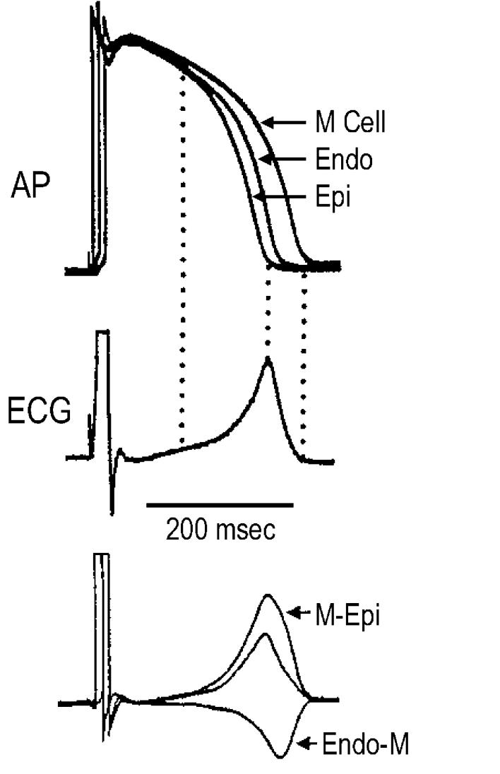
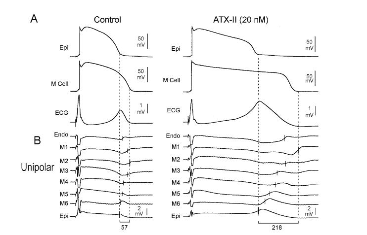
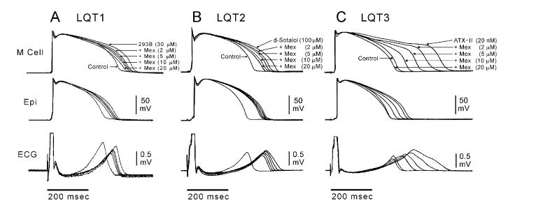
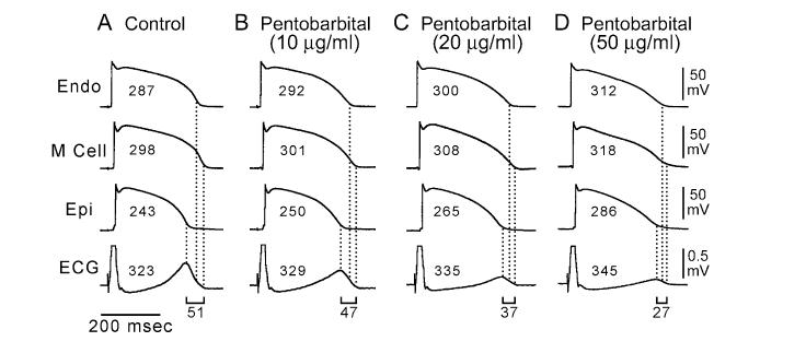
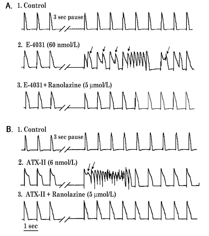
Ventricular myocardium in larger mammals is composed of three distinct cell types: epicardial, M, and endocardial cells. Epicardial and M cell, but not endocardial cell, action potentials have a prominent I(to)-mediated notch. M cells are distinguished from the other cell types in that they display a smaller I(Ks), but a larger late I(Na) and I(Na-Ca). These ionic differences may account for the longer action potential duration (APD) and steeper APD-rate relationship of the M cell. The difference in the time course of repolarization of phase 1 and phase 3 contributes to the inscription of the electrocardiographic J wave and T wave, respectively. These repolarization gradients are modulated by electrotonic interactions, [K(+)](o), and agents or mutations that alter net repolarizing current. An increase in late I(Na), as occurring under a variety of pathophysiological states or in response to certain toxins, leads to a preferential prolongation of the M cell action potential, thus prolonging the QT interval and increasing transmural dispersion of repolarization (TDR), which underlies the development of torsade de pointes (TdP) arrhythmias. Agents that reduce late I(Na) are effective in reducing TDR and suppressing TdP. A reduction in peak I(Na) or an increase in net repolarizing current in the early phases of the action potential can lead to a preferential abbreviation of the action potential of epicardium in the right ventricle, and thus the development of a large TDR, phase 2 reentry, and polymorphic ventricular tachycardia associated with the Brugada syndrome.


Figures

Figure 1.
Figure 1.
Voltage gradients on either side of the M region are responsible for inscription of the electrocardiographic T wave. Top: Action potentials simultaneously recorded from endocardial, epicardial, and M region sites of an arterially perfused canine left ventricular wedge preparation. Middle: ECG recorded across the wedge. Bottom: Computed voltage differences between the epicardium and M region action potentials (Δ VM-Epi) and between the M region and endocardium responses ( ΔVEndo-M). The center trace, the average of the two opposing voltage gradients, closely resembles the ECG. (Modified from Yan and Antzelevitch, with permission.)
Figure 2.
Figure 2.
ATX-II-induced augmentation of late INa amplifies transmural dispersion of repolarization in the coronary-perfused wedge preparation. Each panel shows: A: Transmembrane action potentials recorded from M (M2) and epicardial sites of a canine left ventricular wedge preparation together with a transmural ECG recorded across the bath (BCL of 2,000 ms) in the absence (left) and the presence (right) of ATX-II (20 nmol/L); B: Eight intramural unipolar electrograms recorded approximately 1.2 mm apart from endocardial (Endo), M (6 sites; M1-M6), and epicardial (Epi) regions (120-μm silver electrodes insulated except at the tip) inserted midway into the wedge preparation. Dashed vertical lines in the unipolar electrograms denote the maximum time of the first derivative (Vmax) of the T wave (local repolarization time). (Modified from Antzelevitch et al., with permission.)
Figure 3.
Figure 3.
Block of late INa with mexiletine reduces TDR in experimental models of the long QT syndrome. Each panel shows transmembrane action potentials recorded from M and epicardial (Epi) sites in canine left ventricular wedge preparations together with a transmural ECG recorded across the bath (BCL of 2,000 ms). Traces are recorded in the presence of the IKs blocker, chromanol 293B (LQT1), IKr blocker D-sotalol (LQT2), and late INa agonist, ATX-II (LQT3), plus increasing concentrations of mexiletine. Mexiletine produced a greater abbreviation of the M cell vs epicardial action potential at every concentration, resulting in a reduction in transmural dispersion of repolarization in all three LQTS models. (Modified from Shimizu and Antzelevitch,, with permission.)
Figure 4.
Figure 4.
Pentobarbital-induced QT prolongation but reduction in transmural dispersion of repolarization. Each panel shows transmembrane action potentials recorded from endocardial (Endo), M, and epicardial (Epi) cells in a canine left ventricular wedge preparation together with a transmural ECG recorded across the bath (BCL of 2,000 ms). Numbers in the action potentials denote APD90 values. Numbers above the ECGs denote the QT intervals. (Modified from Shimizu et al., with permission.)
Figure 5.
Figure 5.
Inhibition by ranolazine of pause-triggered early afterdepolarizations (EADs) and ventricular tachycardias (VTs) in the presence of either E-4031 (panel A) or ATX-9II (panel B) in a female rabbit isolated perfused heart paced at 1 Hz. Records 1-3 in each panel were obtained serially from the same heart; panels A and B represent different hearts. Representative monophasic action potentials (MAPs) were recorded before and after a three-second pause during (A1 and B1) control conditions (no drug) and during perfusion with (A2) E-4031 (60 nmol/L) alone, (A3) E-4031 (60 nmol/L) plus ranolazine (5 μmol/L), (B2) ATX-II (6 nmol/L) alone, or (B3) ATX-II (6 nmol/L) plus ranolazine (30 μmol/L). Arrows indicate EADs, EAD-triggered premature ventricular beats, and VTs ranolazine suppressed EADs and VTs induced in the presence of E-4031 (panel A3), and ATX-II (panel B3). Similar results were obtained from five and two additional hearts treated with either E-4031 or ATX-II, respectively (see text).

References

    1. Antzelevitch C, Dumaine R. Electrical heterogeneity in the heart: Physiological, pharmacological and clinical implications. In: Page E, Fozzard HA, Solaro RJ, editors. Handbook of Physiology. Section 2 The Cardiovascular System. Oxford University Press; New York: 2001. pp. 654–692.
    1. Di Diego JM,, Sun ZQ. Antzelevitch C: Ito and action potential notch are smaller in left vs. right canine ventricular epicardium. Am J Physiol. 1996;271:H548–H561. - PubMed
    1. Volders PG, Sipido KR, Carmeliet E, Spatjens RL, Wellens HJ, Vos MA. Repolarizing K+ currents ITO1 and IKs are larger in right than left canine ventricular midmyocardium. Circulation. 1999;99(2):206–210. - PubMed
    1. Sicouri S, Antzelevitch C. A subpopulation of cells with unique electrophysiological properties in the deep subepicardium of the canine ventricle. The M cell. Circ Res. 1991;68:1729–1741. - PubMed
    1. Antzelevitch C, Shimizu W, Yan GX, Sicouri S, Weissenburger J, Nesterenko VV, Burashnikov A, Di Diego JM,, Saffitz JE, Thomas GP. The M cell: Its contribution to the ECG and to normal and abnormal electrical function of the heart. J Cardiovasc Electrophysiol. 1999;10(8):1124–1152. - PubMed

Publication types