. 2006 May 10:3:14.
doi: 10.1186/1742-6413-3-14.

ASC-H in Pap test--definitive categorization of cytomorphological spectrum

Affiliations

ASC-H in Pap test--definitive categorization of cytomorphological spectrum

Mamatha Chivukula et al. Cytojournal. .

Abstract

Objective: The American Society for Colposcopy and Cervical Pathology (ASCCP) guidelines for management of ASC-H is colposcopic examination followed by biopsy. HPV testing (HPVT) is recommended after a negative biopsy result. More definitive interpretation of ASC-H could prevent discomfort and minimize the cost. The purpose of this study was to evaluate association of various cytomorphological patterns of ASC-H with various clinical scenarios.

Methods: We reviewed SurePath (TriPath Imaging, Inc. Burlington, NC, USA) cervical smears interpreted as ASC-H in 161 women (mean age, 37 {15 to 78} years), over 24 months (2002 to 2003). HPVT (Digene, Hybrid Capture II HPV test, Digene Corporation, Gaithersburg, MD, USA) was performed in 20% of cases (33/161) and biopsy results were available in 54 cases (19 with and 35 without HPVT).

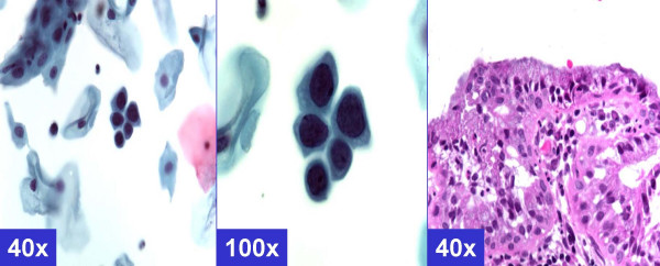
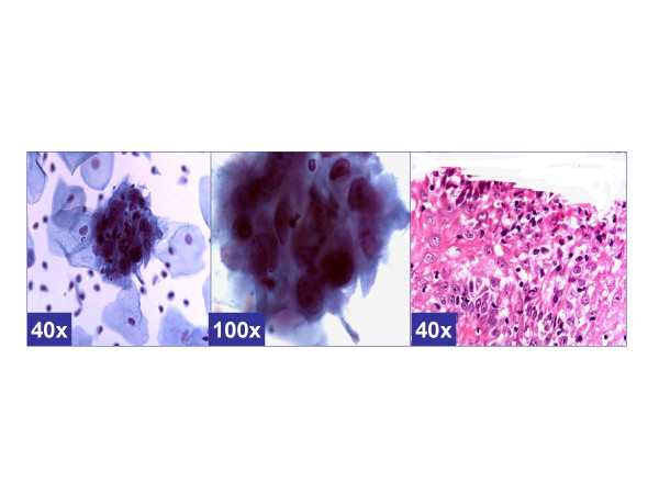
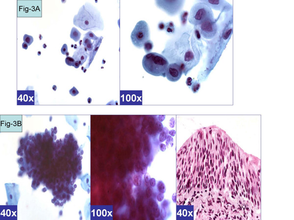
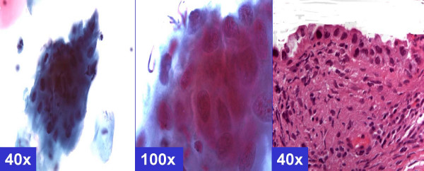
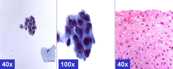
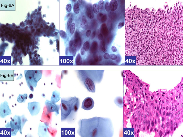
Results: HPVT was positive in 64% (21/33) cases, and negative in 36% (12/33) cases. In the follow-up biopsies of 71% (15/21) of cases with positive HPVT, 27% showed HPV changes or CIN1, 27% showed CIN2-3, and 46% were negative for epithelial abnormality. Follow-up biopsies from cases with negative HPVT (33%, 4/12 cases), 8% showed CIN1 and 25% were negative for any epithelial abnormality. Six cytomorphological patterns of ASC-H correlated with different clinical categories in relation to HPVT and biopsy results. 35% (19 out of 54 ASC-H cases in which biopsy results were available) could be interpreted definitively as HSIL by cytopathology, 11% (6/54) cases as LSIL with cyanophilic atypical parakeratotic pattern, and 31% (17/54) cases as reactive, with HPV status. The interpretation had to be continued as ASC-H in 22% (12/54) cases.

Conclusion: ASC-H demonstrated a spectrum of cytomorphological patterns. Some of these patterns in liquid-based cervical smears may be more specifically interpreted as LSIL, HSIL, or benign if HPV status is known.


Figures

Figure 1
Figure 1
MGH-like pattern (ASC-H, favor reactive) Groups of metaplastic cells arranged in checkerboard like pattern. The dark nuclei may show nucleoli (arrows). (A & b- Cervical smear [Papanicolaou stained SurePath™ Preparation], c- Cervical biopsy [Hematoxylin-eosin stained section]).
Figure 2
Figure 2
Repair-like pattern (ASC-H, favor repair). Cohesive groups of cells with ill-defined school of fish pattern with relatively polarized cells with pointed ends (arrow head) show relatively low N/C ratio. The nuclei show nucleoli (arrows). (a & b- Cervical smear [Papanicolaou stained SurePath™ Preparation], c- Cervical biopsy [Hematoxylin-eosin stained section]).
Figure 3
Figure 3
Atrophy-like pattern (ASC-H, favor atrophy). A. Single cell pattern. Isolated cells with hyperchromatic atypical nuclei with smudgy chromatin). (a & b- Cervical smear [Papanicolaou stained SurePath™ Preparation]). B. HCG of parabasal cells. Cohesive hyperchromatic crowded groups of small parabasal cells with high N/C ratio. The nuclei are relatively small and show nucleoli (arrow). (a & b- Cervical smear [Papanicolaou stained SurePath™ Preparation], c- Cervical biopsy [Hematoxylin-eosin stained section]).
Figure 4
Figure 4
ASC-H: NOS. Cohesive groups of atypical cells with mostly ill-defined cell borders. The nuclei vary in size with coarse chromatin; however, the nuclear details in most are relatively smudgy (arrows). (a & b- Cervical smear [Papanicolaou stained SurePath™ Preparation], c- Cervical biopsy [Hematoxylin-eosin stained section]).
Figure 5
Figure 5
Cyanophilic Atypical parakeratosis (ASC-H, favor HPV). Cohesive groups of hyperchromatic cyanophilic cells with ill-defined cell borders, which are straight with angulations better seen at periphery (arrow). N/C ratio is higher. Chromatin is smudgy. Some cells may show koilocytic space around nuclei. (a & b- Cervical smear [Papanicolaou stained SurePath™ Preparation], c- Cervical biopsy [Hematoxylin-eosin stained section]).
Figure 6
Figure 6
HSIL pattern (ASC-H, favor HSIL). A. Syncytial pattern. Hyperchromatic crowded groups of cells without distinct cell borders. The hyperchromatic nuclei vary in size and show coarsely granular chromatin (arrows). (a & b- Cervical smear [Papanicolaou stained SurePath™ Preparation], c- Cervical biopsy [Hematoxylin-eosin stained section]).B. Singly scattered (so called 'litigation') cells. Scattered, isolated, atypical cells show high N/C ratio. The nuclei have coarse chromatin without nucleoli (arrows). (a & b- Cervical smear [Papanicolaou stained SurePath™ Preparation], c- Cervical biopsy [Hematoxylin-eosin stained section]).

References

    1. Apgar BS, Zoschnick L, Wright TC., Jr The 2001 Bethesda System Terminology. Am FAM Physician. 68:1992–1998. 2003 Nov 15. - PubMed
    1. The Bethesda System Web site Atlas using the 2001 Bethesda System terminology http://www.cytopathology.org/NIH/
    1. Nayar R, Solomon D. Second edition of 'The Bethesda Systemfor reporting cervical cytology' – atlas, website, and Bethesda interobserver reproducibility project. Cytojournal. 1:4. doi: 10.1186/1742-6413-1-4. 2004 Oct 21. - DOI - PMC - PubMed
    1. Garbar C, Mascaux C, Fontaine V. Efficiency of aninexpensive liquid- based cytology performed by cytocentrifugations:a comparative study using the histology as reference standard. Cytojournal. 2:15. doi: 10.1186/1742-6413-2-15. http://www.cytojournal.com/content/2/1/15 2005 Sep 15. - DOI - PMC - PubMed
    1. Shidham VB, Rao RN, Machhi J, Chavan A. Microglandular hyperplasia has a cytomorphological spectrum overlapping with atypical squamous cells-cannot rule out high-grade squamous intraepithelial lesion (ASC-H) Diagn cytopathol. 2004;30:57–61. doi: 10.1002/dc.10342. - DOI - PubMed