. 2006 May 10:3:11.
doi: 10.1186/1742-2094-3-11.

Intracranial administration of deglycosylated C-terminal-specific anti-Abeta antibody efficiently clears amyloid plaques without activating microglia in amyloid-depositing transgenic mice

Affiliations

Intracranial administration of deglycosylated C-terminal-specific anti-Abeta antibody efficiently clears amyloid plaques without activating microglia in amyloid-depositing transgenic mice

Niki C Carty et al. J Neuroinflammation. .

Abstract

Background: Antibodies against the Ass peptide clear Ass deposits when injected intracranially. Deglycosylated antibodies have reduced effector functions compared to their intact counterparts, potentially avoiding immune activation.

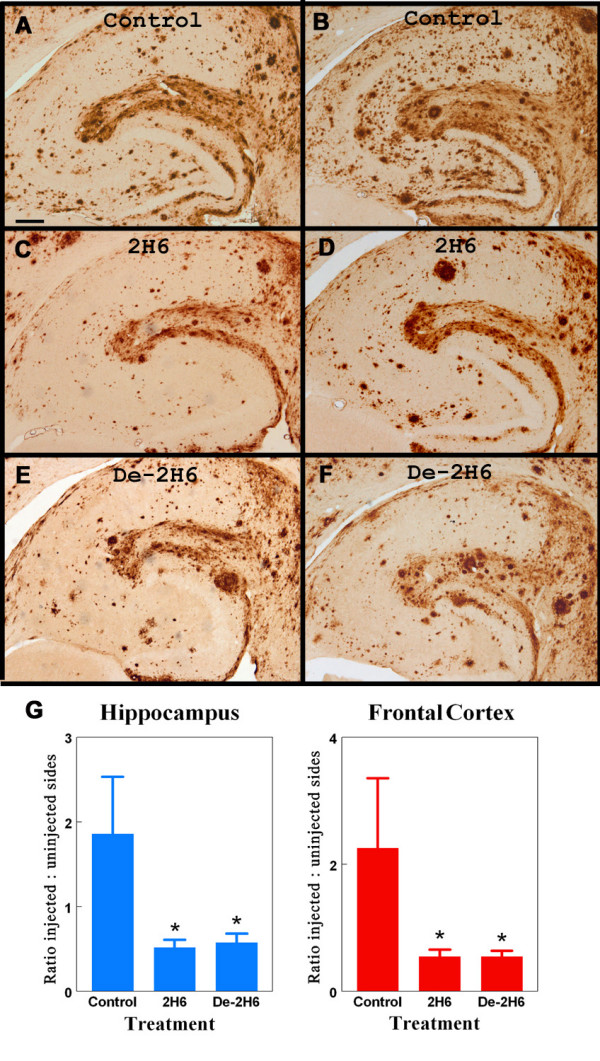
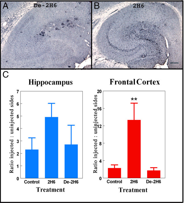
Methods: Deglycosylated or intact C-terminal specific high affinity anti-Abeta antibody (2H6) were intracranially injected into the right frontal cortex and hippocampus of amyloid precursor protein (APP) transgenic mice. The untreated left hemisphere was used to normalize for the extent of amyloid deposition present in each mouse. Control transgenic mice were injected with an antibody against a drosophila-specific protein (amnesiac). Tissues were examined for brain amyloid deposition and microglial responses 3 days after the injection.

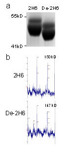
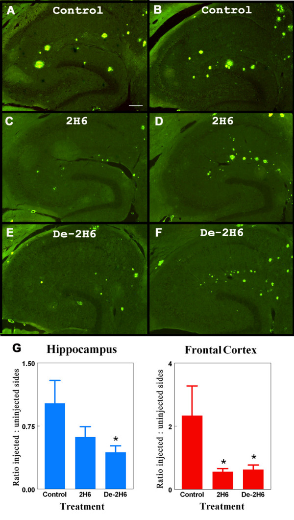
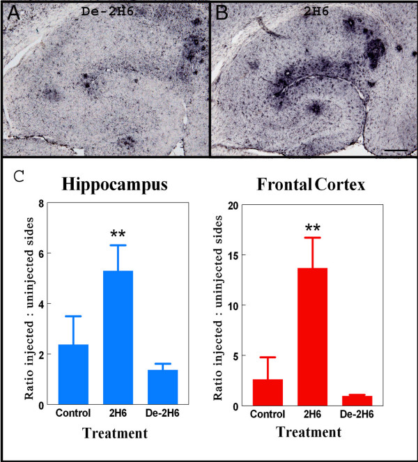
Results: The deglycosylated 2H6 antibody had lower affinity for several murine Fcgamma receptors and human complement than intact 2H6 without a change in affinity for Ass. Immunohistochemistry for Abeta and thioflavine-S staining revealed that both diffuse and compact deposits were reduced by both antibodies. In animals treated with the intact 2H6 antibody, a significant increase in Fcgamma-receptor II/III immunostaining was observed compared to animals treated with the control IgG antibody. No increase in Fcgamma-receptor II/III was found with the deglycosylated 2H6 antibody. Immunostaining for the microglial activation marker CD45 demonstrated a similar trend.

Conclusion: These findings suggest that the deglycosylated 2H6 is capable of removing both compact and diffuse plaques without activating microglia. Thus, antibodies with reduced effector functions may clear amyloid without concomitant immune activation when tested as immunotherapy for Alzheimer's disease.


Figures

Figure 1
Figure 1
Verification of deglycosylation of de-2H6 by and MALDI-TOF-MS and SDS-PAGE. Panel A. SDS-PAGE analysis of 2H6 and de-2H6. Samples were size fractionated under denaturing conditions on a 3–8% Tris-Acetate Gel and stained with Coomassie blue. Note the lower apparent molecular weight for the deglycosylated heavy chain doublet. Panel B. MALDI-TOF-MS analysis revealed the expected 2% reduction in molecular weight after removal of N-linked glycans in the de-2H6 antibody
Figure 2
Figure 2
Total Aß load is reduced following intracranial administration of intact anti-Aβ antibody and deglycosylated anti-Aβ antibody. Panels B, D, and F show total Aβ immunostaining in the left (untreated) hippocampal regions of 20 mo. old APP transgenic mice. Panels A, C, and E show total Aβ staining in right hippocampal regions of 20 mo. old APP transgenic mice receiving intracranial injection of control antibody (panel A) or anti-Aβ C-terminal antibody (2H6; panel C), or deglycosylated anti-Aβ C-terminal antibody (de-2H6; panel E). Magnification = 40×, scale bar = 50 mm. Panel G shows quantification of the Aß load as the ratio of injected (right) side to uninjected (left) side for both the hippocampal and frontal cortical injection sites * indicates P < 0.05 compared to mice injected with control IgG.
Figure 3
Figure 3
Thioflavine S labeled compact amyloid deposits are reduced following intracranial administration of anti-Aβ antibody. Panels B, D, and F show total thioflavine S staining of compact amyloid deposits in left (untreated) hippocampal regions of 20 mo. old APP transgenic mice. Panels A, C, and E show total thioflavine S staining in right hippocampal regions of 20 mo. old APP transgenic mice receiving intracranial injection of control antibody (panel A) or anti-Aβ C-terminal antibody (2H6; panel C), or deglycosylated anti-Aβ C-terminal antibody (de-2H6; panel E). Magnification = 40×, scale bar = 50 μm. Panel G shows quantification of the amyloid load as the ratio of injected (right) side to uninjected (left) side for both the hippocampal and frontal cortical injection sites * indicates P < 0.05 compared to mice injected with control IgG.
Figure 4
Figure 4
Fcγ receptor expression is increased following intracranial administration of intact anti-Aβ antibody but not deglycosylated anti-Aβ antibody. Panels A and B show Fcγ-receptor II/III staining in right hippocampal regions of 20 mo. old APP transgenic mice receiving intracranial injection of deglycosylated C-terminal anti-Aβ antibody (de-2H6) (panel A) or intact anti-Aβ C-terminal antibody (2H6; panel B). Magnification = 40×, scale bar = 50 μm. Panel C shows quantification of the Fcγ-R immunostaining as the ratio of injected (right) side to uninjected (left) side for both the hippocampal and frontal cortical injection sites. ** Indicates P < 0.01 versus both control IgG and de-2H6.
Figure 5
Figure 5
CD45 expression is increased in mice receiving intracranial administration of intact but not deglycosylated anti-Aβ antibody. Panels A and B show total CD45 staining in right hippocampal regions of 20 mo. old APP transgenic mice receiving intracranial injection of deglycosylated C-terminal anti-Aβ antibody (de-2H6) (panel A) or intact anti-Aβ C-terminal antibody (2H6; panel B). Magnification = 40×, scale bar = 50 μm. Panel C shows quantification of theCD45 immunostaining as the ratio of injected (right) side to uninjected (left) side for both the hippocampal and frontal cortical injection sites * indicates P < 0.05 compared to mice injected with control IgG and de-2H6.

References

    1. Bard F, Barbour R, Cannon C, Carretto R, Fox M, Games D, Guido T, Hoenow K, Hu K, Johnson-Wood K, Khan K, Kholodenko D, Lee C, Lee M, Motter R, Nguyen M, Reed A, Schenk D, Tang P, Vasquez N, Seubert P, Yednock T. Epitope and isotype specificities of antibodies to beta -amyloid peptide for protection against Alzheimer's disease-like neuropathology. Proc Natl Acad Sci U S A. 2003;100:2023–2028. doi: 10.1073/pnas.0436286100. - DOI - PMC - PubMed
    1. Masliah E, Hansen L, Adame A, Crews L, Bard F, Lee C, Seubert P, Games D, Kirby L, Schenk D. Abeta vaccination effects on plaque pathology in the absence of encephalitis in Alzheimer disease. Neurology. 2005;64:129–131. - PubMed
    1. Nicoll JA, Wilkinson D, Holmes C, Steart P, Markham H, Weller RO. Neuropathology of human Alzheimer disease after immunization with amyloid-beta peptide: a case report. Nat Med. 2003;9:448–452. doi: 10.1038/nm840. - DOI - PubMed
    1. Orgogozo JM, Gilman S, Dartigues JF, Laurent B, Puel M, Kirby LC, Jouanny P, Dubois B, Eisner L, Flitman S, Michel BF, Boada M, Frank A, Hock C. Subacute meningoencephalitis in a subset of patients with AD after Abeta42 immunization. Neurology. 2003;61:46–54. - PubMed
    1. Cummings BJ, Satou T, Head E, Milgram NW, Cole GM, Savage MJ, Podlisny MB, Selkoe DJ, Siman R, Greenberg BD, Cotman CW. Diffuse plaques contain C-terminal A beta 42 and not A beta 40: evidence from cats and dogs. Neurobiol Aging. 1996;17:653–659. - PubMed