. 2006 May 10;26(19):5248-55.
doi: 10.1523/JNEUROSCI.4483-05.2006.

Synaptic plasticity in CNGA3(-/-) mice: cone bipolar cells react on the missing cone input and form ectopic synapses with rods

Affiliations

Synaptic plasticity in CNGA3(-/-) mice: cone bipolar cells react on the missing cone input and form ectopic synapses with rods

Silke Haverkamp et al. J Neurosci. .

Abstract

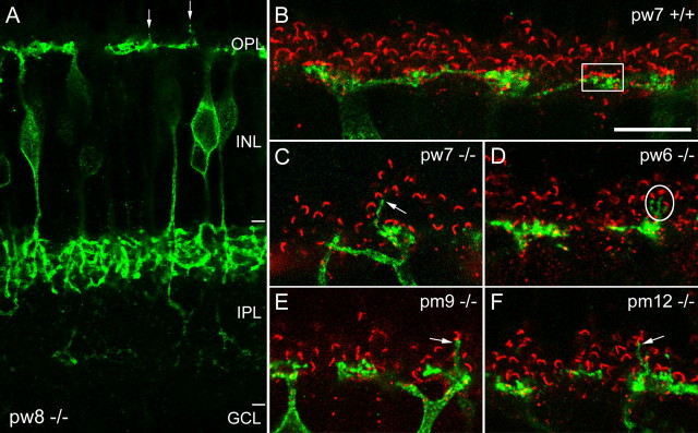
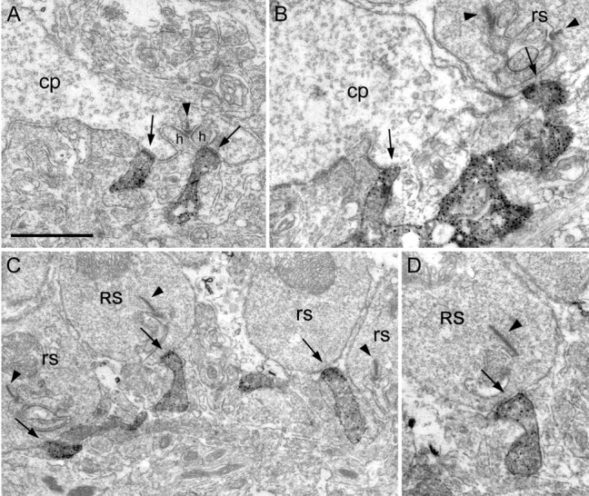
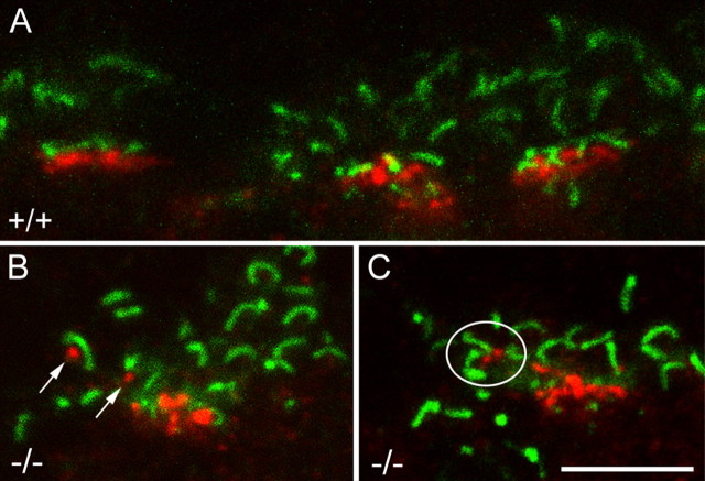
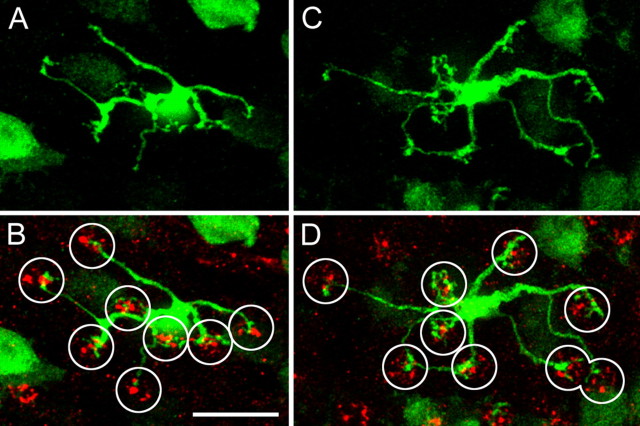
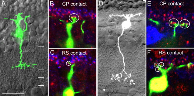
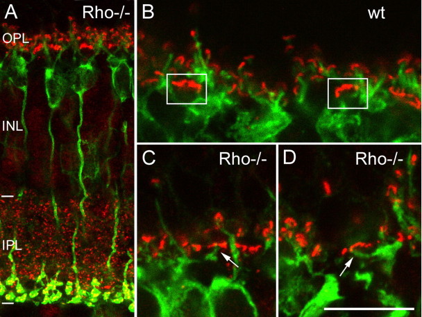
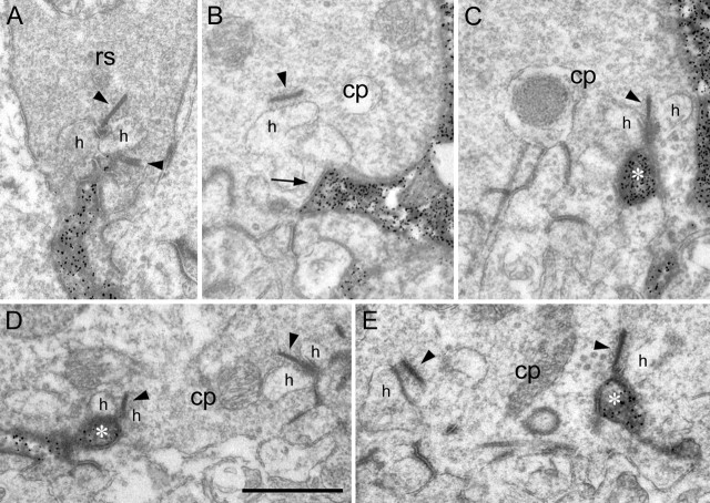
In the mammalian retina, rods and cones connect to distinct sets of bipolar cells. Rods are presynaptic to a single type of rod bipolar cell, whereas cones connect to different types of cone bipolar cells. Synaptic rewiring between cone photoreceptor terminals and rod bipolar cell dendrites has been described as a general result of photoreceptor degeneration. To investigate whether cone bipolar cells also show synaptic plasticity in the absence of cone input, we studied the connectivity of cone bipolar cell dendrites in CNGA3(-/-) mice, a model with specific loss of cone photoreceptor function. Dendritic connections of ON and OFF cone bipolar cells were visualized using specific cell markers or by intracellular injection with fluorescent dyes. The results show that cone bipolar cells in CNGA3(-/-) mice form ectopic synapses with rods. In contrast, cone bipolar cells do not form ectopic synapses with rods in CNGA3(-/-)Rho(-/-) mice, in which both types of photoreceptors are nonfunctional. In analogy with these results, we found that input-deprived rod bipolar cells form ectopic synapses with functional cones in Rho(-/-) mice but not with inoperable cones in the CNGA3(-/-)Rho(-/-) mouse. Our data indicate that the formation of ectopic bipolar cell synapses in the outer plexiform layer requires a functional presynaptic photoreceptor.


Figures

Figure 1.
Figure 1.
Confocal images of NK3R-immunoreactive bipolar cells from vertical sections of CNGA3−/− and wild-type retinas. A, CNGA3−/− mouse retina at pw8. The retinal layers are indicated. INL, Inner nuclear layer; GCL, ganglion cell layer; pm, postnatal month. NK3R immunoreactivity is visible in bipolar cells with axons terminating in the outer IPL. The arrows indicate terminal dendrites, which extend into the outer part of the OPL. B, High-power image of NK3R (green) and bassoon (red) immunoreactivity in the OPL of a wild-type mouse. Bassoon labels the photoreceptor ribbons in rod spherules, where they show a horseshoe-shaped structure, and in cone pedicles, where they are clustered in a row (frame). C–F, NK3R (green) and bassoon (red) immunoreactivity in the OPL of CNGA3−/− mice at the ages indicated. In all cases, NK3R-labeled bipolar cell dendrites extending into the outer part of the OPL can be found. The arrows in C, E, and F mark examples in which the tips of single outgrowing bipolar cell dendrites are in close relationship to bassoon-labeled ribbons, indicating that they are in contact with a rod spherule. The circle in D encloses an example in which three outgrowing dendrites apparently make contact with three rod spherules. Scale bar: (in B) A, 20 μm; B–F, 10 μm.
Figure 2.
Figure 2.
Electron micrographs of a 7-week-old CNGA3−/− mouse retina immunostained with anti-NK3R antibody. A, Cone pedicle (cp) containing one presynaptic ribbon (arrowhead) and two lateral postsynaptic horizontal cell elements (h). Two NK3R-immunoreactive dendrites make flat contacts at the cone pedicle base (arrows). B, Labeling is seen in a process making a flat contact at the cone pedicle base (cp; arrow) and another one onto a rod spherule (rs; arrow). C, Row of rod spherules with potential contacts to NK3R-immunoreactive dendrites (arrows). D, Higher magnification of the second rod spherule (RS) shown in C, but two sections further. Now the flat contact is clearly visible (arrow). Scale bar: (in A) A, B, D, 1 μm; C, 1.43 μm.
Figure 3.
Figure 3.
Confocal images of vertical sections through the outer plexiform layer of wild-type and CNGA3−/− retina double labeled for GluR5 (red) and bassoon (green). A, Wild-type retina with strong punctate immunofluorescence of GluR5 at three cone pedicles. B, C, GluR5 immunoreactivity in the OPL of CNGA3−/− mice. Four examples of red GluR5 puncta (arrows in B, circle in C) at the locations where OFF bipolar cell dendrites contact rod spherules (compare with Fig. 1C–F). Scale bar: (in C) 5 μm.
Figure 4.
Figure 4.
ON cone bipolar cells (type 7) in the GUS–GFP mouse retina. A, C, Horizontal view of two GFP-labeled bipolar cells in a whole-mounted retina (confocal stack from the inner nuclear layer to the OPL; dendritic trees were constructed by collapsing the stacks into a single plane). B, D, Same cells as above double labeled for GFP and for the glutamate receptor subunit GluR5. The clusters of GluR5 puncta represent individual cone pedicles (circles). The dendrites of the type 7 bipolar cell in B contact eight cone pedicles; the cell in D contacts nine cone pedicles. Scale bar: (in B) 10 μm.
Figure 5.
Figure 5.
Confocal images of type 5 and type 7 ON cone bipolar cells in the CNGA3−/− mouse retina. A, Overall morphology of a type 5 ON cone bipolar cell as visualized by projecting a z-stack of optical images and superimposed onto the differential interference contrast image of the corresponding vertical section. The horizontal lines indicate strata S1 through S5 in the IPL. B, Double-labeling immunocytochemistry with antibodies against mGluR6 (red) and kinesin (blue) reveals a contact site with a cone pedicle (CP; circle). C, In a different optical section, a putative synapse with a rod spherule (RS; circle) is visible. D, Overall morphology of a type 7 ON cone bipolar cell. E, In a single optical section, a dendrite of this bipolar cells makes synaptic contacts with two cone pedicles (circles). F, In a different section, three contact sites with rod spherules (circles) are clearly visible. Double-labeling immunocytochemistry same as in B and C. Scale bars: (in A) A, D, 20 μm; B, C, 12 μm; E, F, 10 μm.
Figure 6.
Figure 6.
Vertical sections of Rho−/− and wild-type retinas double labeled for PKCα (green) and CtBP2 (red). A, Low-power image of a Rho−/− mouse retina at pw5. PKCα immunoreactivity is visible in rod bipolar cells with axons terminating deep within the IPL. Antibodies against CtBP2 label all photoreceptor ribbons in the OPL and all bipolar cell ribbons in the IPL. INL, Inner nuclear layer. B, High-power image of PKCα and CtBP2 immunoreactivity in the OPL of a wild-type (wt) mouse. Frames indicate two cone pedicles. PKC-labeled dendrites are in close proximity to CtBP2-labeled ribbons in rod spherules (horseshoe-shaped structure) but not to the labeled ribbons of the two cone pedicles. C, D, OPL of Rho−/− mouse retina at pw5. Two examples of PKC-labeled dendrites with potential contact to cone pedicles (arrows) are shown. Scale bar: (in D) A, 24 μm; B–D, 10 μm.
Figure 7.
Figure 7.
Electron micrographs of PKCα immunoreactivity in the outer plexiform layer of a 5-week-old Rho−/− mouse retina. Synaptic ribbons are marked by arrowheads and horizontal cell processes by “h.” A, Rod spherule (rs) with a presynaptic ribbon, two lateral horizontal cell processes, and a PKCα-labeled invaginating rod bipolar cell dendrite. B, C, Section numbers 1 and 6 of a series of ultrathin sections showing a cone pedicle (cp) with a heavily stained dendrite making flat contact in B (arrow) and invaginating contact in C (asterisk). D, E, Two additional examples of PKCα-immunoreactive dendritic tips making invaginating synaptic contact with the cone pedicle. Scale bar: (in D) 1 μm.

Similar articles

Cited by

References

    1. Biel M, Seeliger M, Pfeifer A, Kohler K, Gerstner A, Ludwig A, Jaissle G, Fauser S, Zrenner E, Hofmann F (1999). Selective loss of cone function in mice lacking the cyclic nucleotide-gated channel CNG3. Proc Natl Acad Sci USA 96:7553–7557. - PMC - PubMed
    1. Brandstätter JH, Fletcher EL, Garner CC, Gundelfinger ED, Wässle H (1999). Differential expression of the presynaptic cytomatrix protein bassoon among ribbon synapses in the mammalian retina. Eur J Neurosci 11:3683–3693. - PubMed
    1. Claes E, Seeliger M, Michalakis S, Biel M, Humphries P, Haverkamp S (2004). Morphological characterization of the retina of the CNGA3−/−Rho−/− mutant mouse lacking functional cones and rods. Invest Ophthalmol Vis Sci 45:2039–2048. - PubMed
    1. Dacheux RF, Raviola E (1986). The rod pathway in the rabbit retina: a depolarizing bipolar and amacrine cell. J Neurosci 6:331–345. - PMC - PubMed
    1. Daniele LL, Lillo C, Lyubarsky AL, Nikonov SS, Philp N, Mears AJ, Swaroop A, Williams DS, Pugh EN Jr (2005). Cone-like morphological, molecular, and electrophysiological features of the photoreceptors of the Nrl knockout mouse. Invest Ophthalmol Vis Sci 46:2156–2167. - PMC - PubMed

Publication types

MeSH terms

Substances