

Case Reports
Diffusion-weighted MR imaging characteristics of an acute strokelike form of multiple sclerosis
Affiliations
- PMID: 16687533
- PMCID: PMC7975747
Item in Clipboard
Case Reports
Diffusion-weighted MR imaging characteristics of an acute strokelike form of multiple sclerosis
AJNR Am J Neuroradiol.
2006 May.
Abstract
The characteristics of multiple sclerosis (MS) lesions on diffusion-weighted sequences and apparent diffusion coefficient (ADC) mapping at the very early phase of symptoms have not been clearly described. We report the case of a young woman who presented with a sudden pseudostroke form of MS resulting in hemiplegia and sudden aphasia. MR imaging showed a lesion of the left internal capsule with reduced ADC, which suggests an ischemic stroke. This case shows that very acute MS lesions may have reduced ADC on MR imaging, reflecting cytotoxic and not vasogenic edema.
Figures
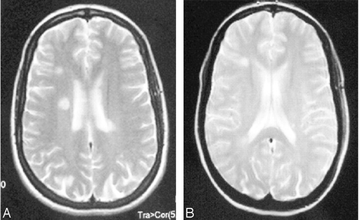
Brain MR imaging, T2-weighted sequence: high-intensity white matter images, 2 of which are periventricular.
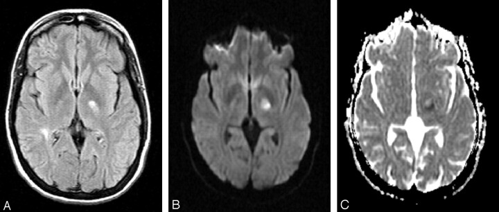
Brain MR imaging 2 hours after onset of symptoms. A, FLAIR sequence, showing high signal intensity of left internal capsule. B, DWI, showing high signal intensity of left internal capsule. C, ADC mapping, showing reduced ADC of the left internal capsule lesion.
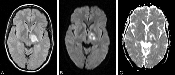
Brain MR imaging on day 10. A, FLAIR sequence, showing extension of the left internal capsule lesion. B, DWI, showing extension of the left internal capsule lesion. C, ADC mapping, showing heterogeneous ADC of the left internal capsule lesion.
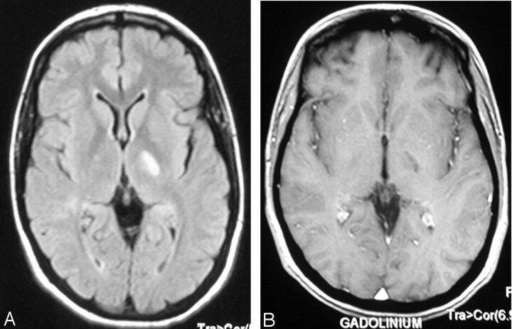
Brain MR imaging at 3 months. A, FLAIR sequence, showing regression of the high signal intensity of the left internal capsule. B, Gadolinium-enhanced T1-weighted image, showing no contrast enhancement of the lesion.
Similar articles
-
Acute multiple sclerosis lesion: conversion of restricted diffusion due to vasogenic edema.J Neuroimaging. 2011 Apr;21(2):202-4. doi: 10.1111/j.1552-6569.2009.00443.x. J Neuroimaging. 2011. PMID: 19888931 Free PMC article.
-
Use of perfusion- and diffusion-weighted imaging in differential diagnosis of acute and chronic ischemic stroke and multiple sclerosis.Neurol Res. 2008 Oct;30(8):816-26. doi: 10.1179/174313208X341003. Neurol Res. 2008. PMID: 18826808
-
Temporal evolution of acute multiple sclerosis lesions on serial sodium (23Na) MRI.Mult Scler Relat Disord. 2019 Apr;29:48-54. doi: 10.1016/j.msard.2019.01.027. Epub 2019 Jan 17. Mult Scler Relat Disord. 2019. PMID: 30669020
-
Diffusion-weighted MR of the brain: methodology and clinical application.Radiol Med. 2005 Mar;109(3):155-97. Radiol Med. 2005. PMID: 15775887 Review. English, Italian.
-
[The clinical application of diffusion weighted magnetic resonance imaging to acute cerebrovascular disorders].No To Shinkei. 1998 Sep;50(9):787-95. No To Shinkei. 1998. PMID: 9789301 Review. Japanese.
Cited by
-
Use of gadolinium-based contrast agents in multiple sclerosis: a review by the ESMRMB-GREC and ESNR Multiple Sclerosis Working Group.Eur Radiol. 2024 Mar;34(3):1726-1735. doi: 10.1007/s00330-023-10151-y. Epub 2023 Sep 2. Eur Radiol. 2024. PMID: 37658891 Review.
-
Comparison of diffusion-weighted imaging and contrast-enhanced T1-weighted imaging on a single baseline MRI for demonstrating dissemination in time in multiple sclerosis.BMC Neurol. 2014 May 7;14:100. doi: 10.1186/1471-2377-14-100. BMC Neurol. 2014. PMID: 24885357 Free PMC article.
-
Acute demyelinating lesions with restricted diffusion in multiple sclerosis.Mult Scler. 2012 Dec;18(12):1745-53. doi: 10.1177/1352458512445407. Epub 2012 Apr 20. Mult Scler. 2012. PMID: 22523157 Free PMC article.
-
Is It Possible to Discriminate Active MS Lesions with Diffusion Weighted Imaging?Eurasian J Med. 2019 Oct;51(3):219-223. doi: 10.5152/eurasianjmed.2019.18473. Eurasian J Med. 2019. PMID: 31692763 Free PMC article.
-
Diffusion-weighted imaging in acute demyelinating myelopathy.Neuroradiology. 2012 Jun;54(6):573-8. doi: 10.1007/s00234-011-0907-6. Epub 2011 Jul 9. Neuroradiology. 2012. PMID: 21743997
References
-
- Devere TR, Trotter JL, Cross AH. Acute aphasia in multiple sclerosis. Arch Neurol 2000;57:1207–09 - PubMed
-
- Lacour A, De Seze J, Revenco E, et al. Acute aphasia in multiple sclerosis: a multicenter study of 22 patients. Neurology 2004;62:974–77 - PubMed
-
- Horsfield MA. Using diffusion-weighted MRI in multicenter clinical trials for multiple sclerosis. J Neurol Sci 2001;186 (suppl 1):S51–4 - PubMed
-
- Castriota-Scanderberg A, Sabatini U, Fasano F, et al. Diffusion of water in demyelinating lesions. Neuroradiology 2002;44:764–67 - PubMed
Publication types
MeSH terms
LinkOut - more resources
Full Text Sources
Medical