

MR Imaging of BK virus encephalitis
- PMID: 16687535
- PMCID: PMC7975713
MR Imaging of BK virus encephalitis
Abstract
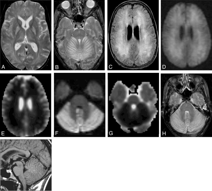
BK virus infection is most often associated with urologic disease in patients who have undergone renal or bone marrow transplantation. We report a rare case of biopsy-confirmed BK virus encephalitis in an immunocompromised patient with hemorrhagic cystitis, in whom dramatic imaging findings were present despite relatively mild clinical symptoms. MR imaging demonstrated widespread increased signal intensity on T2- and fluid-attenuated inversion recovery-weighted images, with restricted diffusion, in the cerebellum, cerebral white matter, and deep gray matter structures. The simultaneous presence of urologic abnormalities and neurologic deficits in certain immunocompromised patients should prompt consideration of BK virus encephalitis.
Figures
Similar articles
-
Rare presentation of BK encephalitis in a child: imaging and pathological findings.Pediatr Radiol. 2012 Sep;42(9):1145-8. doi: 10.1007/s00247-012-2369-3. Epub 2012 Mar 20. Pediatr Radiol. 2012. PMID: 22430483
-
BK virus encephalitis in an immunocompetent patient.Arch Neurol. 1996 Jan;53(1):101-3. doi: 10.1001/archneur.1996.00550010121025. Arch Neurol. 1996. PMID: 8599551
-
BKV-DNA and JCV-DNA in CSF of patients with suspected meningitis or encephalitis.Infection. 2003 Dec;31(6):374-8. doi: 10.1007/s15010-003-3078-5. Infection. 2003. PMID: 14735377
-
BK virus in solid organ transplant recipients: an emerging syndrome.Transplantation. 2001 Nov 27;72(10):1587-92. doi: 10.1097/00007890-200111270-00001. Transplantation. 2001. PMID: 11726814 Review.
-
BK virus-associated nephropathy with hydronephrosis in a patient with AIDS: a case report and literature review.Clin Nephrol. 2016 Mar;85(3):173-8. doi: 10.5414/CN108482. Clin Nephrol. 2016. PMID: 26249547 Review.
Cited by
-
BK virus encephalopathy and sclerosing vasculopathy in a patient with hypohidrotic ectodermal dysplasia and immunodeficiency.Acta Neuropathol Commun. 2016 Jul 13;4(1):73. doi: 10.1186/s40478-016-0342-3. Acta Neuropathol Commun. 2016. PMID: 27411570 Free PMC article.
-
Rare presentation of BK encephalitis in a child: imaging and pathological findings.Pediatr Radiol. 2012 Sep;42(9):1145-8. doi: 10.1007/s00247-012-2369-3. Epub 2012 Mar 20. Pediatr Radiol. 2012. PMID: 22430483
-
BK virus encephalitis without concurrent hemorrhagic cystitis in an allogeneic hematopoietic stem cell transplant recipient.Blood Res. 2013 Sep;48(3):226-8. doi: 10.5045/br.2013.48.3.226. Epub 2013 Sep 25. Blood Res. 2013. PMID: 24086945 Free PMC article. No abstract available.
-
BK nephropathy in the native kidneys of patients with organ transplants: Clinical spectrum of BK infection.World J Transplant. 2016 Sep 24;6(3):472-504. doi: 10.5500/wjt.v6.i3.472. World J Transplant. 2016. PMID: 27683628 Free PMC article. Review.
-
Different behaviour of BK-virus infection in liver transplant recipients.World J Gastroenterol. 2016 Jan 28;22(4):1532-40. doi: 10.3748/wjg.v22.i4.1532. World J Gastroenterol. 2016. PMID: 26819520 Free PMC article. Review.
References
-
- Reploeg MD, Storch GA, Clifford DB. BK virus: a clinical review. Clin Infect Dis 2001;33:191–202 - PubMed
-
- Shinohara T, Matsuda M, Cheng SH, et al. BK virus infection of the human urinary tract. J Med Virol 1993;41:301–05 - PubMed
-
- Hogan TF, Broden EC, McBain JA, et al. Human polymovirus infections with JC virus and BK virus in renal transplant patients. Ann Intern Med 1980;92:373–78 - PubMed
-
- Elsner C, Dorries K. Evidence of human polyomavirus BK and JC infection in normal brain tissue. Virology 1992;191:72–80 - PubMed
Publication types
MeSH terms
LinkOut - more resources
Full Text Sources
Medical