

Diffusion anisotropy and diffusivity of white matter tracts within the temporal stem in Alzheimer disease: evaluation of the "tract of interest" by diffusion tensor tractography
- PMID: 16687540
- PMCID: PMC7975725
Diffusion anisotropy and diffusivity of white matter tracts within the temporal stem in Alzheimer disease: evaluation of the "tract of interest" by diffusion tensor tractography
Abstract
Purpose: Our aim was to determine whether diffusion anisotropy and diffusivity of white matter tracts of the temporal stem in patients with Alzheimer (AD) can be evaluated independently by using diffusion tensor tractography.
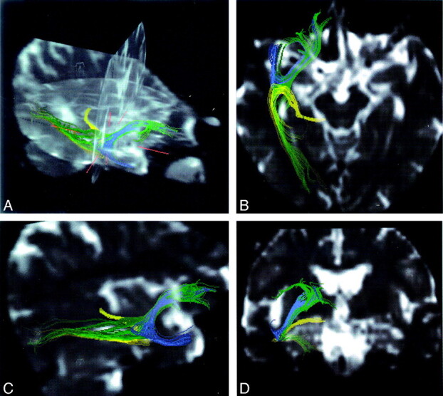
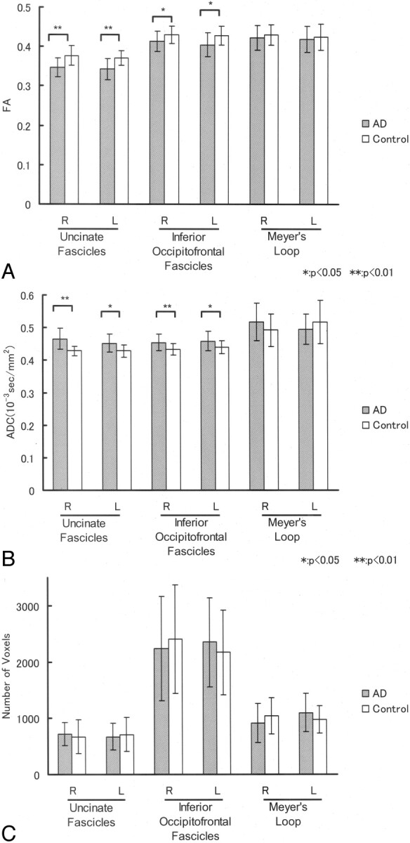
Materials and methods: Subjects included 15 patients with AD (11 women and 4 men; mean age, 74 years) and 15 age-matched control subjects (11 women and 4 men; mean age, 72 years). Diffusion tensor images were acquired by using echo-planar imaging. We drew tractographies of the uncinate fasciculus, inferior occipitofrontal fasciculus, and Meyer's loop, with diffusion tensor analysis software. We measured diffusion anisotropy, diffusivity, and the number of voxels along the "tracts of interest" and used the Student t test to compare results between patients with AD and controls.
Results: Values of diffusion anisotropy of the bilateral uncinate fasciculus and left inferior occipitofrontal fasciculus were significantly lower for patients with AD than for controls. Also, values of diffusivity in the bilateral uncinate fasciculus were significantly greater for patients with AD than for controls. There was no significant difference in diffusion anisotropy or diffusivity along Meyer's loop between the 2 groups. There was no significant difference in the number of voxels included in all constructed tracts between patients with AD and controls.
Conclusion: White matter tracts of the temporal stem can be evaluated independently by using diffusion tensor tractography, which appears to be a promising technique for determining changes in white matter in degenerative diseases.
Figures
Similar articles
-
Fractional anisotropy--threshold dependence in tract-based diffusion tensor analysis: evaluation of the uncinate fasciculus in Alzheimer disease.AJNR Am J Neuroradiol. 2009 Oct;30(9):1700-3. doi: 10.3174/ajnr.A1698. Epub 2009 Jun 18. AJNR Am J Neuroradiol. 2009. PMID: 19541775 Free PMC article.
-
Uncinate fasciculus-correlated cognition in Alzheimer's disease: a diffusion tensor imaging study by tractography.Psychogeriatrics. 2010 Mar;10(1):15-20. doi: 10.1111/j.1479-8301.2010.00312.x. Psychogeriatrics. 2010. PMID: 20594282
-
Diffusion abnormalities of the uncinate fasciculus in Alzheimer's disease: diffusion tensor tract-specific analysis using a new method to measure the core of the tract.Neuroradiology. 2008 Apr;50(4):293-9. doi: 10.1007/s00234-007-0353-7. Epub 2008 Feb 2. Neuroradiology. 2008. PMID: 18246334 Clinical Trial.
-
[Magnetic resonance diffusion tractography in the brain--its application and limitation].Brain Nerve. 2007 May;59(5):467-76. Brain Nerve. 2007. PMID: 17533972 Review. Japanese.
-
Tract-specific analysis for investigation of Alzheimer disease: a brief review.Jpn J Radiol. 2010 Aug;28(7):494-501. doi: 10.1007/s11604-010-0460-y. Epub 2010 Aug 27. Jpn J Radiol. 2010. PMID: 20799014 Review.
Cited by
-
Diffusion tensor imaging of normal-appearing white matter in mild cognitive impairment and early Alzheimer disease: preliminary evidence of axonal degeneration in the temporal lobe.AJNR Am J Neuroradiol. 2007 Nov-Dec;28(10):1943-8. doi: 10.3174/ajnr.A0700. Epub 2007 Sep 28. AJNR Am J Neuroradiol. 2007. PMID: 17905894 Free PMC article.
-
Asymmetry, sex differences and age-related changes in the white matter in the healthy elderly: a tract-based study.BMC Res Notes. 2011 Oct 4;4:378. doi: 10.1186/1756-0500-4-378. BMC Res Notes. 2011. PMID: 21970546 Free PMC article.
-
Cerebral white matter damage in frontotemporal dementia assessed by diffusion tensor tractography.Neuroradiology. 2008 Jul;50(7):605-11. doi: 10.1007/s00234-008-0379-5. Epub 2008 Apr 1. Neuroradiology. 2008. PMID: 18379765 Clinical Trial.
-
Long Longitudinal Tract Lesion Contributes to the Progression of Alzheimer's Disease.Front Neurol. 2020 Oct 16;11:503235. doi: 10.3389/fneur.2020.503235. eCollection 2020. Front Neurol. 2020. PMID: 33178095 Free PMC article.
-
Feasibility of 1.6-mm isotropic voxel diffusion tensor tractography in depicting limbic fibers.Neuroradiology. 2008 Feb;50(2):131-6. doi: 10.1007/s00234-007-0317-y. Epub 2007 Oct 16. Neuroradiology. 2008. PMID: 17938897
References
-
- Ebeling U, von Cramon D. Topography of the uncinate fascicle and adjacent temporal fiber tracts. Acta Neurochir (Wien) 1992;115:143–48 - PubMed
-
- Hanyu H, Sakurai H, Iwamoto T, et al. Diffusion-weighted MR imaging of the hippocampus and temporal white matter in Alzheimer’s disease. J Neurol Sci 1998;156:195–200 - PubMed
-
- Masutani Y, Aoki S, Abe O, et al. MR diffusion tensor imaging: recent advance and new techniques for diffusion tensor visualization. Eur J Radiol 2003;46:53–66 - PubMed
MeSH terms
LinkOut - more resources
Full Text Sources
Medical