

Degree of hippocampal atrophy is related to side of seizure onset in temporal lobe epilepsy
- PMID: 16687541
- PMCID: PMC7975739
Degree of hippocampal atrophy is related to side of seizure onset in temporal lobe epilepsy
Abstract
Background and purpose: Temporal lobe epilepsy (TLE) is associated with pathologic changes in hippocampal physiology and morphology. Our aim was to quantify volume reduction of the right and left hippocampus in patients with TLE and to investigate whether the degree of hippocampal atrophy is related to the side of seizure onset.
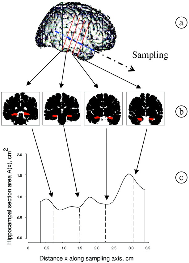
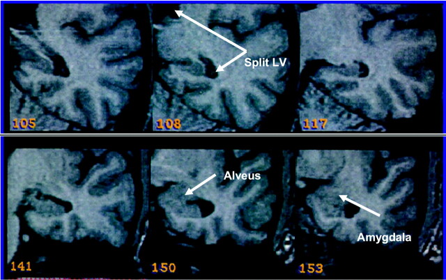
Methods: The volume of the right and left hippocampus was estimated for 50 controls and 101 patients with TLE, by applying the unbiased Cavalieri method on MR images.
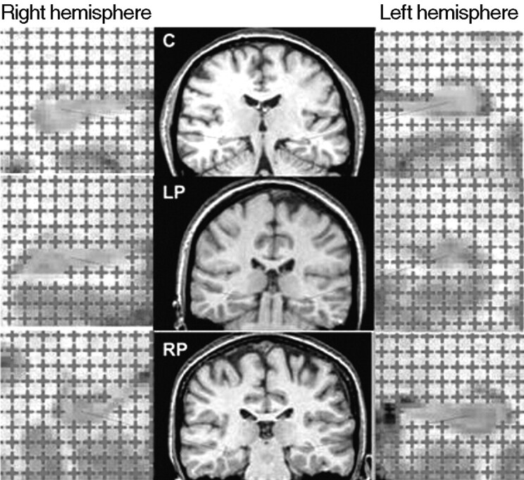
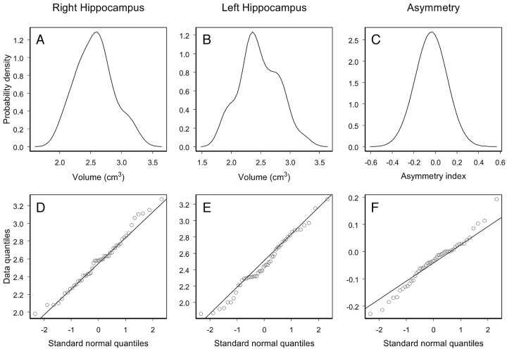
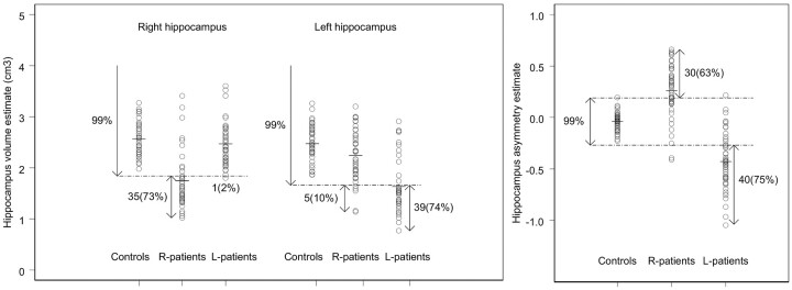
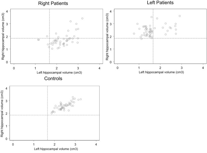
Results: Pairwise comparisons, within a multivariate analysis of variance and adjusted by using the Bonferroni correction, revealed that both right and left hippocampal volumes were, on average, significantly smaller in patients with right-sided seizure onset (R-patients) relative to those of controls (P < .001 and P = .04, respectively). Furthermore, left hippocampal volume was significantly smaller in patients with left-sided seizure onset (L-patients) compared with controls (P < .001), but the right-sided hippocampal volume was not significantly smaller (P = .71). Moreover, a correlation analysis revealed that the strong linear association between the right and left hippocampal volumes existing in the control population (r = 0.73) is partially lost in patients with TLE (r < or = 0.48), and this loss in correlation appears to be more pronounced in L-patients than in R-patients.
Conclusion: Our MR imaging results suggest that although the major damage in patients with TLE is located in the hippocampus ipsilateral to the side of seizure onset, R-patients are more likely to have bilateral hippocampal volume reduction. These findings support the hypothesis that cerebral hemispheres may not only differ in their functionality organization but also in their vulnerability to a neurologic insult.
Figures
References
-
- Norman RM, Sandry S, Corsellis JAN. The nature and origin of patho-anatomical change in the epileptic brain. In: Vinken PJ, Bruyn GW, eds. Handbook of Clinical Neurology, Vol. 15. Amsterdam, Holland: North Holland Publishing Company;1974. :611–20
-
- Duncan JS. Imaging and epilepsy. Brain 1997;120:339–77 - PubMed
-
- Babb TL, Brown WJ. Pathological findings in epilepsy. In: Engel J, ed. Surgical Treatment of the Epilepsies. New York: Raven Press;1987. :511–40
-
- Cendes F, Andermann F, Gloor P, et al. MRI volumetric measurement of amygdala and hippocampus in temporal lobe epilepsy. Neurology 1993;43:719–25 - PubMed
-
- Cook MJ, Fish DR, Shorvon SD, et al. Hippocampal volumetric and morphometric studies in frontal and temporal lobe epilepsy. Brain 1992;115:1001–15 - PubMed
Publication types
MeSH terms
LinkOut - more resources
Full Text Sources