. 2006 Jul;17(7):3329-44.
doi: 10.1091/mbc.e05-12-1146. Epub 2006 May 10.

Adenovirus E4orf4 hijacks rho GTPase-dependent actin dynamics to kill cells: a role for endosome-associated actin assembly

Affiliations

Adenovirus E4orf4 hijacks rho GTPase-dependent actin dynamics to kill cells: a role for endosome-associated actin assembly

Amélie Robert et al. Mol Biol Cell. 2006 Jul.

Abstract

The adenovirus early region 4 ORF4 protein (E4orf4) triggers a novel death program that bypasses classical apoptotic pathways in human cancer cells. Deregulation of the cell cytoskeleton is a hallmark of E4orf4 killing that relies on Src family kinases and E4orf4 phosphorylation. However, the cytoskeletal targets of E4orf4 and their role in the death process are unknown. Here, we show that E4orf4 translocates to cytoplasmic sites and triggers the assembly of a peculiar juxtanuclear actin-myosin network that drives polarized blebbing and nuclear shrinkage. We found that E4orf4 activates the myosin II motor and triggers de novo actin polymerization in the perinuclear region, promoting endosomes recruitment to the sites of actin assembly. E4orf4-induced actin dynamics requires interaction with Src family kinases and involves a spatial regulation of the Rho GTPases pathways Cdc42/N-Wasp, RhoA/Rho kinase, and Rac1, which make distinct contributions. Remarkably, activation of the Rho GTPases is required for induction of apoptotic-like cell death. Furthermore, inhibition of actin dynamics per se dramatically impairs E4orf4 killing. This work provides strong support for a causal role for endosome-associated actin dynamics in E4orf4 killing and in the regulation of cancer cell fate.


Figures
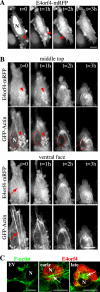
Figure 1.
Figure 1.
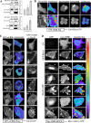
The cytoplasmic translocation of E4orf4 is associated with dynamic changes in actin. (A and B) MCF7 cells were transfected with E4orf4-mRFP alone (A) or with GFP-Actin (B). Twenty-four hours after transfection (t = 0), cells were treated with 75 μM cycloheximide and were observed by time-lapse microscopy for a 3-h period at 10-min intervals. (A) Arrowheads indicate the cytoplasmic translocation of E4orf4. (B) The microscope was refocused to allow changes in actin at the ventral face and in the middle-top part of the cell to be followed simultaneously. Key frames from the two focal planes recorded at the indicated time after 24 h of E4orf4 expression are shown. Red dashed lines delineate the nucleus revealed by labeling with cell-permeable Hoechst, which condenses as the cell retracts and enters the blebbing phase (2 h). Arrowheads designate the juxtanuclear network of GFP-actin-coated vesicles enriched in E4orf4-mRFP and arrows show stress fibers at the cell ventral face. (C) Confocal images of 293T transfected with the vector only (EV) or with Flag-E4orf4 after immunostaining of E4orf4 (anti-2420) and F-actin (phalloidin). Arrows, perinuclear actin ring. N, nucleus. Bars, 10 μm.
Figure 2.
Figure 2.
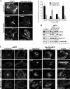
E4orf4 induces myosin II activation in juxtanuclear regions and polarized blebbing. (A) Confocal images of mono-p-MLC staining (p-MLC [Ser19]) in 293T transfected with the vector-only (EV) counterstained with fluorescent phalloidin (F-actin), compared with cells transfected with E4orf4-mRFP in the early stages of E4orf4 expression (early) and in the late blebbing stage (late). Arrowheads indicate p-MLC accumulation in juxtanuclear regions. The white dashed line delineates the cell nucleus. (B) 293T cells were transfected with the vector only (EV) or with mutant E4orf4 constructs displaying alanine substitutions on the indicated residues: E4orf4, wild type; 6R-A, mutant defective in Src binding; R81/F84A and F84A, mutants defective in PP2A binding. Equal amounts of total cell extracts were analyzed by Western blot using anti-p-MLC (Ser19) and anti-p-MLC (Thr18/Ser19). The levels of E4orf4 proteins were revealed using anti-Flag (M2) and β-actin levels are shown as loading controls. Right, 293T were treated with the Src family kinase inhibitor SU6656 (5 μM) or with the vehicle (dimethyl sulfoxide [DMSO]) during and after transfection and p-MLC was analyzed by Western blot 24 h after transfection. (C) Confocal images of F-actin (phalloidin) and myosin heavy chain IIb (anti-MHC IIb) in MCF7 transfected with the vector, compared with cells expressing E4orf4-mRFP, in cells treated with DMSO versus 50 μM blebbistatin for 1 h before cell fixation. Arrowhead shows the colocalization of F-actin and MHC IIb in E4orf4 cell, which are organized into a juxtanuclear contractile ring from which prominent stress fibers radiate to the cell periphery. The white dashed lines delineate the cell nucleus (N). Bars, 10 μm. Graph shows the ratios of the average intensity of E4orf4-mRFP in the nucleus over that in the cytoplasm in E4orf4 cells treated with DMSO versus blebbistatin (***p < 0.0001). Data are the means ± SD of at least 30 cells (n). (D) Crude cell fractionation of control (EV) versus cells transfected with Flag-E4orf4. Equal amounts of cellular fractions were analyzed by Western blot using anti-p-MLC (Ser19), anti-histone H3 (nuclei), anti-GM130 (Golgi membranes), anti-TOM20 (mitochondria) and anti-Flag (E4orf4). P1, nuclei and tightly associated membranes; P2, heavy membranes; P3, light membranes; C, cytosol. CytoD, cells were treated with cytoD for 30 min before cell fractionation. W/O, without cytoD treatment. (E) Phase contrast micrographs from time-lapse analyses of the blebbing dynamics of E4orf4-expressing 293T cells, compared with 293T treated with nocodazole (100 ng/ml; 16 h). Arrows show the direction of the protruding/retracting blebs and the white dashed lines indicate the polarized axes of blebbing. See Supplemental Video 1.
Figure 3.
Figure 3.
E4orf4 induces de novo actin polymerization in the perinuclear region. MCF7 or 293T cells were transfected as indicated and fixed after a 5-min incubation with fluorescent G-actin in saponin-permeabilization buffer. Where indicated, 5 μM cytoD was added during the in situ polymerization assay. (A) Epifluorescence images of MCF7 transfected with the vector (EV) or E4orf4-mRFP and fixed after G-actin incorporation. The nuclei were labeled with Hoechst, and the remaining E4orf4-mRFP staining in the nucleus and the cytoplasm (insoluble) is shown in red. Dashed circle lines show examples of the juxtanuclear regions used for the quantitative analyses presented in B. (B) Average intensity of pixels was measured over the entire cell or in the juxtanuclear regions from the original confocal images using the MetaMorph software version 4.5 (***p < 0.0001, **p < 0.001, and *p < 0.01). Data are the means ± SD of 15–58 cells (n) from at least three independent experiments. E4orf4(6R-A), mutant defective in Src binding; R81/F84A, mutant defective in PP2A binding. (C) Confocal images of 293T transfected with EV (top) or E4orf4-GFP (bottom) and fixed after a 5-min G-actin incorporation. F-actin staining was performed after cell fixation using fluorescent-labeled phalloidin to compare the preexisting actin network with the newly formed dynamics microfilaments that incorporate G-actin. Arrows and arrowheads indicate dynamic sites of actin nucleation at the juxtanuclear ring displaying insoluble E4orf4 protein. N, nucleus. Bars, 10 μm.
Figure 4.
Figure 4.
E4orf4-induced actin nucleation is not associated with Golgi membranes. (A) MCF7 or HeLa cells transfected with the Golgi marker GalT-GFP alone (EV) or with E4orf4-mRFP were fixed after a 5-min incubation with Alexa-647–labeled G-actin in saponin-permeabilization buffer and analyzed by confocal microscopy. Representative images of HeLa cells show the juxtanuclear position of the Golgi complex (EV) and the absence of overlap between Golgi membranes (GalT-GFP) and the dynamic juxtanuclear actin ring in E4orf4 expressing cell. (B) Transfected HeLa cells were incubated with DMSO or 5 μg/ml BFA to disrupt the Golgi complex, either before E4orf4 expression for a 4-h period (chronic treatment) or after the onset of actin assembly for a 30-min period (acute treatment), followed by in situ G-actin incorporation. Confocal images and quantification of de novo actin polymerization show that BFA treatment effectively disrupts the Golgi complex, without interfering with the formation or maintenance of the actin ring. Juxtanuclear actin nucleation was quantified by measuring the average intensity of pixels from the original cross-section images using the MetaMorph software version 4.5. Data are the means ± SD of at least eight individual cells (n). ***p < 0.0001. Dashed lines delineate the nucleus (N). Bars, 10 μm.
Figure 5.
Figure 5.
E4orf4 promotes the nucleation of dynamic actin particles associated with endosomes motility. Confocal images of MCF7 transfected with the vector (EV) or E4orf4-mRFP and fixed after incorporation of Alexa-647–labeled transferrin for 45 min, followed by a 5-min incorporation of Alexa-488–labeled G-actin. Arrows indicate the region in insets showing high magnifications of the asymmetric association of endosomal vesicles and the tubular-vesicular actin structures in juxtanuclear regions. Note that endosomes were massively recruited to the dynamic actin ring. (B) Confocal images of MCF7 transfected with E4orf4-mRFP and GFP-GPI, stained with Alexa-647–labeled phalloidin (F-actin) after cell fixation. High magnification of the juxtanuclear region show the clear overlapping patterns of the GPI-positive endosomes with F-actin tails. (C) Multiphoton time-lapse imaging of GFP-actin in MCF7 cells transfected with E4orf4-mRFP and GFP-actin. The time series at 30-s intervals show the association of GFP-actin comet tails with large vesicles (arrowheads) that fused with one another. Note that some vesicles were totally covered with GFP-actin (arrow) and some others were associated with trailing actin tails (arrowhead). (D) Confocal time-lapse imaging of GFP-actin and RFP-Rab11 in cells expressing Flag-E4orf4, showing the recruitment of Rab11-positive endosomes to the forming actin network. High magnifications of the region delineated by the white box show dynamics vesicular structures decorated with RFP-Rab11 and associated with small tails of GFP-actin (arrow). These motile vesicles were recruited to the perinuclear actin fibers and displayed a rocket-like movement reminiscent of actin comets (see Supplemental Video 2). The red track in the last panel indicates the movement of the pointed vesicle over a 48-s period (arrow). Dual channel acquisitions were performed at an interval of 2 s/frame. Bars, 10 μm.
Figure 6.
Figure 6.
Spatial activation of Rho proteins by E4orf4. (A) 293T cells were transfected with the indicated plasmids DNA, and pull-down assays were performed using GST-pCRIB 24 h after transfection. The bound material was analyzed by Western blot and levels of the GTPases and Flag-E4orf4 in total lysates are shown (TL). The relative levels of active GTPases were estimated by densitometric analyses and are the means ± SD of three independent experiments. (B and C) Confocal or multiphoton images of live cells transfected with the indicated plasmids DNA, together with YFP-pCRIB (B) or with GFP-wCRIB (C) showing representative recruitment patterns of the reporter probes for active Cdc42 and Rac. (D) Cells expressing the indicated constructs together with the reporter probes for active Rho proteins myc-rRBD were analyzed by epifluorescence microscopy after cell fixation followed by myc-rRBD staining using anti-myc (9E10). Where appropriate, a pseudocolor intensity scale was used to highlight the relative amount of membrane-associated CRIB or RBD, over the total amount of CRIB or RBD in individual cells (from black to white: low-to-high intensity). Arrows and arrowheads designate membrane vesicles and cortical protrusions, respectively, in which E4orf4 recruits the CRIB and RBD probes, respectively. Activation of Rho proteins is impaired in cells expressing the mutant E4orf4 (6R-A) defective in Src binding, as revealed by the lack of CRIB and RBD recruitment, but not in cells expressing the mutant E4orf4 (R81/F84A) defective in PP2A binding. Bars, 10 μm.
Figure 7.
Figure 7.
Distinct contributions of Rho proteins to E4orf4-induced de novo actin polymerization. (A) Top, confocal images of N-Wasp-GFP in MCF7 cells coexpressing myc-Cdc42, after a 45-min treatment with 50 μM wiskostatin (N-Wasp inhibitor) or DMSO. Middle and bottom, confocal images showing two focal planes of the same MCF7 cells transfected with E4orf4-mRFP. Cells were fixed after a 45-min incubation with or without wiskostatin, followed by a 5-min incorporation of fluorescent G-actin. Note that inhibition of N-Wasp blocks actin polymerization at the juxtanuclear actin ring (arrows) but not in stress fibers spanning the ventral face of the same cells. Graph, cells were treated for 45-min with DMSO, wiskostatin, 100 μM NSC23766 (Rac1 inhibitor), or 10 μM Y-27632 (ROCK inhibitor) before G-actin incorporation. G-actin incorporation was quantified in the juxtanuclear region versus the entire ventral face of the cells by measuring the average intensity of pixels in each regions from the original confocal cross section and ventral face images of the same cell, using the MetaMorph software version 4.5 (right). Data are the means ± SD of at least 10 individual cells (n). (B) Western blots of total cell lysates from control and MCF7 cells transfected with 75 nM siRNAs to GFP, Cdc42, RhoA, or Rac1 48 h posttransfection. The levels of Rho proteins were revealed using the indicated antibodies and calreticulin protein levels are shown as loading control. Titration curves were established by loading increasing amounts of the control extract (25, 10, and 5 μg) to estimate the percentage of inhibition of Cdc42, RhoA, and Rac1, -2 and -3 protein levels (italic numbers) in extracts from siRNA-transfected cells (25 μg) by densitometric analyses. Note that the antibody against Rac1, -2 and -3 revealed >55% reduction of Rac isoforms, suggesting a potent reduction of Rac1. (C) Confocal images of MCF7 cells transfected with the indicated siRNAs and mRFP (left) or transfected with the siRNAs and E4orf4-mRFP. Seventy-two hours after siRNA transfection (24 h after E4orf4-mRFP transfection), cells were processed for labeling with fluorescent transferrin followed by G-actin incorporation. Cross sections and ventral face images of the same cells show representative phenotypes observed at the level of juxtanuclear actin nucleation and endosomes recruitment, and nucleation of stress fibers formation, respectively. The percentages of cells with actin structures showing endosome-associated actin vesicles are indicated and were estimated from two independent experiments, n > 25 cells. Bars, 10 μm.
Figure 8.
Figure 8.
RhoA, but not Cdc42 or Rac1, is required for E4orf4-induced myosin II activation. (A) 293T cells transfected with E4orf4 were incubated with DMSO or with the Rho kinase inhibitor (10 μM Y-27632) for a 2-h period 24 h after transfection or with the Rac1 inhibitor (100 μM NSC23766) added 6 h after transfection for a 18-h period (middle), or cells were transfected with the indicated siRNAs to Rho proteins 48 h before E4orf4 transfection (right). Cells were lysed 24 h after E4orf4 transfection, and equal amounts of cell extracts were analyzed by Western blot using the indicated antibodies. (B) Confocal images of MCF7 cells transfected with the indicated siRNAs 48 h before the transfection with Flag-E4orf4 or Flag-E4orf4-GFP or treated with the Rac1 inhibitor (100 μM NSC23766) for 16 h before E4orf4 transfection. Cells were fixed 24 h after E4orf4 transfection and processed for double immunostaining of p-MLC (Ser19) and F-actin (phalloidin). The focal plan displaying the highest p-MLC staining is shown. Flag-E4orf4 was immunolabeled using the anti-E4orf4 (2419) antibody. Arrows show p-MLC-containing stress fibers in E4orf4 cells transfected with siRNAs to Cdc42. Arrowheads indicate the filopodia-like microfilaments, which accumulate in E4orf4 cells transfected with siRNAs to RhoA or Rac1 or were treated with the Rac1 inhibitor. Bar, 10 μm.
Figure 9.
Figure 9.
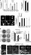
E4orf4-induced actin dynamics trigger apoptotic-like cell death and cell killing. (A) 293T or MCF7 cells were transfected with Flag-E4orf4 alone or together with the indicated siRNAs or plasmid DNA to inhibit Cdc42 (CRIB-N-Wasp), Rho (RBD-Rhotekin), Rho kinase (ROCK2 CAT KD), Arp2/3 complex (Scar VCA), or myosin II (MLC-AA), or treated with the myosin II inhibitor blebbistatin (293T, 10 μM; MCF7, 50 μM), or the Rac1 inhibitor NSC23766 (100 μM). Cells were fixed 24 h after transfection with Flag-E4orf4 and processed for immunostaining of both E4orf4 and the interfering proteins, followed by DAPI labeling to visualize the nuclear morphology. The nuclear condensation (apoptotic-like nuclear shrinkage and chromatin condensation) was determined by counting the number of cotransfected cells with apoptotic-like condensation and is expressed as the percentage of inhibition relative to cells expressing E4orf4 alone that displayed at least 35 to 50% of the cell population with apoptotic-like nuclei (inset graph). Data are the mean ± SD of three independent experiments, n > 1000. Micrographs at the bottom left show an example of E4orf4-induced nuclear condensation, which is inhibited in cells coexpressing the Scar VCA. Bar, 10 μM. Top right, the nuclear condensation was evaluated in live cells transfected with mRFP (EV) or E4orf4-mRFP together with histone H2A-GFP and treated with vehicle (DMSO) or blebbistatin immediately after transfection, n > 100. (B) Colony-forming assays. 293T cells were transfected with pGKpuro together with the vector only (EV) or Flag-E4orf4 using a plasmid DNA ratio of 1:25. Other cultures were cotransfected with a nonphosphorylatable mutant MLC (AA). Where indicated, blebbistatin (1.5–3.0 μM) was added to the culture medium immediately after transfection and replaced every 72 h. Aliquots of transfected cells were kept for Western blot analyses of expression levels 24h after transfection (middle, inset), before applying the selection for transfected cells (3 μg/ml puromycin) with or without blebbistatin for 12 d. Percentages of surviving cells were obtained by counting the number of resulting colonies (top) and are expressed relative to the total number of colonies obtained in cells transfected with the vector only and similarly processed. Data are the means ± SD of 3 independent experiments. Bottom left, immunostaining of E4orf4 in the surviving colonies showing high levels of E4orf4 only in blebbistatin-treated cells. Middle and right, double immunostaining with anti-E4orf4 and anti-Ki67 (cell proliferation marker) of a E4orf4-positive colony, compared with a E4orf4-negative colony in samples treated with blebbistatin. E4orf4-positive cells proliferate, but to a lower rate, as revealed by a twofold decrease in the size of colonies. Bar, 30 μm.
Figure 10.
Figure 10.
Working models. (A) Three-dimensional representation of the peculiar structure of the juxtanuclear actin network, showing the spatial contributions of Rho proteins to E4orf4-induced actin dynamics (see Discussion for details). We speculate that the nucleation of actin comets at the surface of endosomes (dark gray circles) drives their recruitment to the juxtanuclear region where they provide a localized input of microfilaments and actin modifiers for assembly of the contractile ring. (B) E4orf4 hijacks Rho GTPases signaling to kill cells in a way that requires interaction with Src-family kinases. Potential mechanisms include the activation of RhoGEFs and/or inhibition of RhoGAPs through Src-mediated phosphorylation. We propose that E4orf4-induced actin dynamics lead to severe perturbations of vesicular traffic and organelle-based membrane dynamics, which trigger the activation of an endo-lysosomal–based caspase-independent death pathway.

Similar articles

Cited by

References

    1. Amano M., Chihara K., Kimura K., Fukata Y., Nakamura N., Matsuura Y., Kaibuchi K. Formation of actin stress fibers and focal adhesions enhanced by Rho-kinase. Science. 1997;275:1308–1311. - PubMed
    1. Avalos A. M., Arthur W. T., Schneider P., Quest A. F., Burridge K., Leyton L. Aggregation of integrins and RhoA activation are required for Thy-1-induced morphological changes in astrocytes. J. Biol. Chem. 2004;279:39139–39145. - PubMed
    1. Bagrodia S., Taylor S. J., Jordon K. A., Van Aelst L., Cerione R. A. A novel regulator of p21-activated kinases. J. Biol. Chem. 1998;273:23633–23636. - PubMed
    1. Baum B., Kunda P. Actin nucleation: spire-actin nucleator in a class of its own. Curr. Biol. 2005;15:R305–R308. - PubMed
    1. Bement W. M., Benink H. A., von Dassow G. A microtubule-dependent zone of active RhoA during cleavage plane specification. J. Cell Biol. 2005;170:91–101. - PMC - PubMed

Publication types

MeSH terms